4AQD

Crystal structure of fully glycosylated human butyrylcholinesterase


Experimental Data Snapshot

  • Method: X-RAY DIFFRACTION
  • Resolution: 2.50 Å
  • R-Value Free: 0.232 
  • R-Value Work: 0.163 
  • R-Value Observed: 0.165 

wwPDB Validation   3D Report Full Report


Ligand Structure Quality Assessment 


This is version 2.1 of the entry. See complete history


Literature

Human butyrylcholinesterase produced in insect cells: huprine-based affinity purification and crystal structure.

Brazzolotto, X.Wandhammer, M.Ronco, C.Trovaslet, M.Jean, L.Lockridge, O.Renard, P.Y.Nachon, F.

(2012) FEBS J 279: 2905-2916

  • DOI: https://doi.org/10.1111/j.1742-4658.2012.08672.x
  • Primary Citation of Related Structures:  
    4AQD

  • PubMed Abstract: 

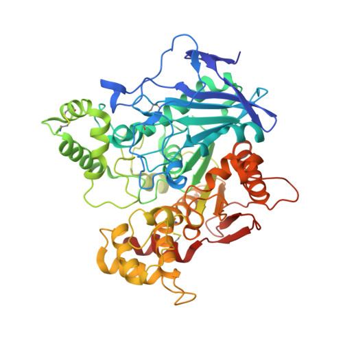
    Butyrylcholinesterase (BChE) is a serine hydrolase that is present in all mammalian tissues. It can accommodate larger substrates or inhibitors than acetylcholinesterase (AChE), the enzyme responsible for hydrolysis of the neurotransmitter acetylcholine in the central nervous system and neuromuscular junctions. AChE is the specific target of organophosphorous pesticides and warfare nerve agents, and BChE is a stoichiometric bioscavenger. Conversion of BChE into a catalytic bioscavenger by rational design or designing reactivators specific to BChE required structural data obtained using a recombinant low-glycosylated human BChE expressed in Chinese hamster ovary cells. This expression system yields ≈ 1 mg of pure enzyme per litre of cell culture. Here, we report an improved expression system using insect cells with a fourfold higher yield for truncated human BChE with all glycosylation sites present. We developed a fast purification protocol for the recombinant protein using huprine-based affinity chromatography, which is superior to the classical procainamide-based affinity. The purified BChE crystallized under different conditions and space group than the recombinant low-glycosylated protein produced in Chinese hamster ovary cells. The crystals diffracted to 2.5 Å. The overall monomer structure is similar to the low-glycosylated structure except for the presence of the additional glycans. Remarkably, the carboxylic acid molecule systematically bound to the catalytic serine in the low-glycosylated structure is also present in this new structure, despite the different expression system, purification protocol and crystallization conditions.


  • Organizational Affiliation

    Département de Toxicologie, Institut de Recherche Biomédicale des Armées, La Tronche, France. xavier@brazzolotto.net


Macromolecules
Find similar proteins by:  (by identity cutoff)  |  3D Structure
Entity ID: 1
MoleculeChains Sequence LengthOrganismDetailsImage
BUTYRYLCHOLINESTERASE
A, B
531Homo sapiensMutation(s): 0 
EC: 3.1.1.8
UniProt & NIH Common Fund Data Resources
Find proteins for P06276 (Homo sapiens)
Explore P06276 
Go to UniProtKB:  P06276
PHAROS:  P06276
GTEx:  ENSG00000114200 
Entity Groups  
Sequence Clusters30% Identity50% Identity70% Identity90% Identity95% Identity100% Identity
UniProt GroupP06276
Sequence Annotations
Expand
  • Reference Sequence
Oligosaccharides

Help

Entity ID: 2
MoleculeChains Length2D Diagram Glycosylation3D Interactions
2-acetamido-2-deoxy-beta-D-glucopyranose-(1-4)-2-acetamido-2-deoxy-beta-D-glucopyranose
C, E
2N-Glycosylation
Glycosylation Resources
GlyTouCan:  G42666HT
GlyCosmos:  G42666HT
GlyGen:  G42666HT
Entity ID: 3
MoleculeChains Length2D Diagram Glycosylation3D Interactions
2-acetamido-2-deoxy-beta-D-glucopyranose-(1-4)-[beta-L-fucopyranose-(1-6)]2-acetamido-2-deoxy-beta-D-glucopyranose
D, F, H, I, J
D, F, H, I, J, K
3N-Glycosylation
Glycosylation Resources
GlyTouCan:  G28454KX
GlyCosmos:  G28454KX
GlyGen:  G28454KX
Entity ID: 4
MoleculeChains Length2D Diagram Glycosylation3D Interactions
alpha-D-mannopyranose-(1-4)-2-acetamido-2-deoxy-beta-D-glucopyranose-(1-4)-2-acetamido-2-deoxy-beta-D-glucopyranose
G
3N-Glycosylation
Glycosylation Resources
GlyTouCan:  G62182OO
GlyCosmos:  G62182OO
GlyGen:  G62182OO
Small Molecules
Ligands 8 Unique
IDChains Name / Formula / InChI Key2D Diagram3D Interactions
NAG
Query on NAG

Download Ideal Coordinates CCD File 
IA [auth B]
JA [auth B]
KA [auth B]
LA [auth B]
M [auth A]
IA [auth B],
JA [auth B],
KA [auth B],
LA [auth B],
M [auth A],
MA [auth B]
2-acetamido-2-deoxy-beta-D-glucopyranose
C8 H15 N O6
OVRNDRQMDRJTHS-FMDGEEDCSA-N
PG4
Query on PG4

Download Ideal Coordinates CCD File 
N [auth A],
O [auth A]
TETRAETHYLENE GLYCOL
C8 H18 O5
UWHCKJMYHZGTIT-UHFFFAOYSA-N
PEG
Query on PEG

Download Ideal Coordinates CCD File 
NA [auth B],
OA [auth B]
DI(HYDROXYETHYL)ETHER
C4 H10 O3
MTHSVFCYNBDYFN-UHFFFAOYSA-N
BAL
Query on BAL

Download Ideal Coordinates CCD File 
HA [auth B],
L [auth A]
BETA-ALANINE
C3 H7 N O2
UCMIRNVEIXFBKS-UHFFFAOYSA-N
GLY
Query on GLY

Download Ideal Coordinates CCD File 
GA [auth A],
IB [auth B]
GLYCINE
C2 H5 N O2
DHMQDGOQFOQNFH-UHFFFAOYSA-N
EDO
Query on EDO

Download Ideal Coordinates CCD File 
P [auth A]
PA [auth B]
Q [auth A]
QA [auth B]
R [auth A]
P [auth A],
PA [auth B],
Q [auth A],
QA [auth B],
R [auth A],
RA [auth B],
S [auth A],
SA [auth B],
T [auth A],
TA [auth B],
U [auth A],
UA [auth B],
V [auth A],
VA [auth B],
W [auth A],
WA [auth B],
X [auth A],
XA [auth B],
YA [auth B]
1,2-ETHANEDIOL
C2 H6 O2
LYCAIKOWRPUZTN-UHFFFAOYSA-N
CL
Query on CL

Download Ideal Coordinates CCD File 
CA [auth A]
DA [auth A]
DB [auth B]
EA [auth A]
EB [auth B]
CA [auth A],
DA [auth A],
DB [auth B],
EA [auth A],
EB [auth B],
GB [auth B],
HB [auth B]
CHLORIDE ION
Cl
VEXZGXHMUGYJMC-UHFFFAOYSA-M
UNX
Query on UNX

Download Ideal Coordinates CCD File 
AA [auth A]
AB [auth B]
BA [auth A]
BB [auth B]
CB [auth B]
AA [auth A],
AB [auth B],
BA [auth A],
BB [auth B],
CB [auth B],
FA [auth A],
FB [auth B],
Y [auth A],
Z [auth A],
ZA [auth B]
UNKNOWN ATOM OR ION
X
Experimental Data & Validation

Experimental Data

  • Method: X-RAY DIFFRACTION
  • Resolution: 2.50 Å
  • R-Value Free: 0.232 
  • R-Value Work: 0.163 
  • R-Value Observed: 0.165 
  • Space Group: P 21 21 21
Unit Cell:
Length ( Å )Angle ( ˚ )
a = 72.75α = 90
b = 79.26β = 90
c = 227.2γ = 90
Software Package:
Software NamePurpose
PHENIXrefinement
XDSdata reduction
XSCALEdata scaling
PHENIXphasing

Structure Validation

View Full Validation Report


Ligand Structure Quality Assessment 


Entry History 

Deposition Data

Revision History  (Full details and data files)

  • Version 1.0: 2012-07-04
    Type: Initial release
  • Version 1.1: 2012-08-08
    Changes: Database references
  • Version 1.2: 2013-11-20
    Changes: Derived calculations
  • Version 1.3: 2018-11-14
    Changes: Advisory, Data collection, Database references
  • Version 1.4: 2019-03-06
    Changes: Data collection, Derived calculations, Experimental preparation
  • Version 2.0: 2020-07-29
    Type: Remediation
    Reason: Carbohydrate remediation
    Changes: Advisory, Atomic model, Data collection, Derived calculations, Other, Structure summary
  • Version 2.1: 2023-12-20
    Changes: Advisory, Data collection, Database references, Derived calculations, Refinement description, Structure summary