4APU

PR X-Ray structures in agonist conformations reveal two different mechanisms for partial agonism in 11beta-substituted steroids


Experimental Data Snapshot

  • Method: X-RAY DIFFRACTION
  • Resolution: 1.90 Å
  • R-Value Free: 0.252 
  • R-Value Work: 0.205 
  • R-Value Observed: 0.208 

wwPDB Validation   3D Report Full Report


Ligand Structure Quality Assessment 


This is version 1.3 of the entry. See complete history


Literature

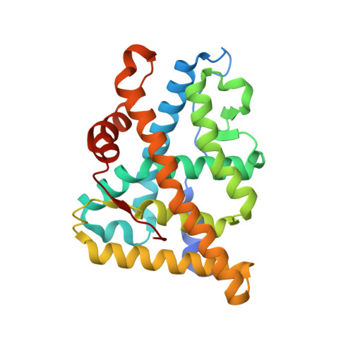
X-ray structures of progesterone receptor ligand binding domain in its agonist state reveal differing mechanisms for mixed profiles of 11 beta-substituted steroids.

Lusher, S.J.Raaijmakers, H.C.Vu-Pham, D.Kazemier, B.Bosch, R.McGuire, R.Azevedo, R.Hamersma, H.Dechering, K.Oubrie, A.van Duin, M.de Vlieg, J.

(2012) J Biol Chem 287: 20333-20343

  • DOI: https://doi.org/10.1074/jbc.M111.308403
  • Primary Citation of Related Structures:  
    4A2J, 4APU

  • PubMed Abstract: 

    We present here the x-ray structures of the progesterone receptor (PR) in complex with two mixed profile PR modulators whose functional activity results from two differing molecular mechanisms. The structure of Asoprisnil bound to the agonist state of PR demonstrates the contribution of the ligand to increasing stability of the agonist conformation of helix-12 via a specific hydrogen-bond network including Glu(723). This interaction is absent when the full antagonist, RU486, binds to PR. Combined with a previously reported structure of Asoprisnil bound to the antagonist state of the receptor, this structure extends our understanding of the complex molecular interactions underlying the mixed agonist/antagonist profile of the compound. In addition, we present the structure of PR in its agonist conformation bound to the mixed profile compound Org3H whose reduced antagonistic activity and increased agonistic activity compared with reference antagonists is due to an induced fit around Trp(755), resulting in a decreased steric clash with Met(909) but inducing a new internal clash with Val(912) in helix-12. This structure also explains the previously published observation that 16α attachments to RU486 analogs induce mixed profiles by altering the binding of 11β substituents. Together these structures further our understanding of the steric and electrostatic factors that contribute to the function of steroid receptor modulators, providing valuable insight for future compound design.


  • Organizational Affiliation

    Departments of Molecular Design and Informatics, MSD, P. O. Box 20, 5340 BH, Oss, The Netherlands.s.lusher@esciencecenter.nl


Macromolecules
Find similar proteins by:  (by identity cutoff)  |  3D Structure
Entity ID: 1
MoleculeChains Sequence LengthOrganismDetailsImage
PROGESTERONE RECEPTOR
A, B
260Homo sapiensMutation(s): 0 
UniProt & NIH Common Fund Data Resources
Find proteins for P06401 (Homo sapiens)
Explore P06401 
Go to UniProtKB:  P06401
PHAROS:  P06401
GTEx:  ENSG00000082175 
Entity Groups  
Sequence Clusters30% Identity50% Identity70% Identity90% Identity95% Identity100% Identity
UniProt GroupP06401
Sequence Annotations
Expand
  • Reference Sequence
Small Molecules
Ligands 3 Unique
IDChains Name / Formula / InChI Key2D Diagram3D Interactions
A2K
Query on A2K

Download Ideal Coordinates CCD File 
E [auth B](8S,11R,13S,14S,16S,17S)-17-cyclopropylcarbonyl-16-ethenyl-13-methyl-11-(4-pyridin-3-ylphenyl)-2,6,7,8,11,12,14,15,16,17-decahydro-1H-cyclopenta[a]phenanthren-3-one
C35 H37 N O2
AUWGCRAJBXNPNU-WWVPXYRRSA-N
OR8
Query on OR8

Download Ideal Coordinates CCD File 
C [auth A]2-CHLORO-N-[[4-(3,5-DIMETHYLISOXAZOL-4-YL)PHENYL]METHYL]-1,4-DIMETHYL-1H-PYRAZOLE-4-SULFONAMIDE
C17 H19 Cl N4 O3 S
XNTNHEPWXKNVBH-UHFFFAOYSA-N
SO4
Query on SO4

Download Ideal Coordinates CCD File 
D [auth A]SULFATE ION
O4 S
QAOWNCQODCNURD-UHFFFAOYSA-L
Binding Affinity Annotations 
IDSourceBinding Affinity
A2K BindingDB:  4APU IC50: 0.61 (nM) from 1 assay(s)
EC50: 0.66 (nM) from 1 assay(s)
Experimental Data & Validation

Experimental Data

  • Method: X-RAY DIFFRACTION
  • Resolution: 1.90 Å
  • R-Value Free: 0.252 
  • R-Value Work: 0.205 
  • R-Value Observed: 0.208 
  • Space Group: P 1 21 1
Unit Cell:
Length ( Å )Angle ( ˚ )
a = 57.662α = 90
b = 64.374β = 96.33
c = 70.468γ = 90
Software Package:
Software NamePurpose
REFMACrefinement
d*TREKdata reduction
SCALAdata scaling
REFMACphasing

Structure Validation

View Full Validation Report


Ligand Structure Quality Assessment 


Entry History 

Deposition Data

Revision History  (Full details and data files)

  • Version 1.0: 2012-04-25
    Type: Initial release
  • Version 1.1: 2012-07-04
    Changes: Other
  • Version 1.2: 2018-02-07
    Changes: Database references, Experimental preparation
  • Version 1.3: 2023-12-20
    Changes: Data collection, Database references, Derived calculations, Other, Refinement description