4AMC

Crystal structure of Lactobacillus reuteri 121 N-terminally truncated glucansucrase GTFA


Experimental Data Snapshot

  • Method: X-RAY DIFFRACTION
  • Resolution: 3.60 Å
  • R-Value Free: 0.260 
  • R-Value Work: 0.239 
  • R-Value Observed: 0.240 

wwPDB Validation   3D Report Full Report


This is version 1.3 of the entry. See complete history


Literature

Structure of the Alpha-1,6/Alpha-1,4-Specific Glucansucrase Gtfa from Lactobacillus Reuteri 121

Pijning, T.Vujicic-Zagar, A.Kralj, S.Dijkhuizen, L.Dijkstra, B.W.

(2012) Acta Crystallogr Sect F Struct Biol Cryst Commun 68: 1448

  • DOI: https://doi.org/10.1107/S1744309112044168
  • Primary Citation of Related Structures:  
    4AMC

  • PubMed Abstract: 

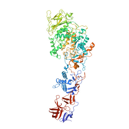
    The reuteransucrase GTFA from Lactobacillus reuteri 121, which belongs to glycosyl hydrolase family GH70, synthesizes branched α-glucans with both α-1,6- and α-1,4-glycosidic linkages (reuteran) from sucrose. The crystal structure of GTFA-ΔN, a 118 kDa fragment of GTFA comprising residues 745-1763 and including the catalytic domain, was determined at 3.6 Å resolution by molecular replacement. The crystals have large solvent channels and an unusually high solvent content of 85%. GTFA-ΔN has the same domain arrangement and domain topologies as observed in previously determined GH70 glucansucrase structures. The architecture of the GTFA-ΔN active site and binding pocket confirms that glucansucrases have a conserved substrate specificity for sucrose. However, this first crystal structure of an α-1,6/α-1,4-specific glucansucrase shows that residues from conserved sequence motif IV (1128-1136 in GTFA-ΔN) contribute to the acceptor-binding subsites and that they display differences compared with other structurally characterized glucansucrases. In particular, the structure clarifies the importance of residues following the transition-state stabilizer for product specificity, and especially residue Asn1134, which is in a position to interact with sugar units in acceptor subsite +2.


  • Organizational Affiliation

    Laboratory of Biophysical Chemistry, University of Groningen, Nijenborgh 7, 9747 AG Groningen, The Netherlands.


Macromolecules
Find similar proteins by:  (by identity cutoff)  |  3D Structure
Entity ID: 1
MoleculeChains Sequence LengthOrganismDetailsImage
GLUCANSUCRASE1,049Limosilactobacillus reuteriMutation(s): 0 
EC: 2.4.1.5
UniProt
Find proteins for Q5SBL9 (Limosilactobacillus reuteri)
Explore Q5SBL9 
Go to UniProtKB:  Q5SBL9
Entity Groups  
Sequence Clusters30% Identity50% Identity70% Identity90% Identity95% Identity100% Identity
UniProt GroupQ5SBL9
Sequence Annotations
Expand
  • Reference Sequence
Small Molecules
Ligands 1 Unique
IDChains Name / Formula / InChI Key2D Diagram3D Interactions
CA
Query on CA

Download Ideal Coordinates CCD File 
B [auth A]CALCIUM ION
Ca
BHPQYMZQTOCNFJ-UHFFFAOYSA-N
Experimental Data & Validation

Experimental Data

  • Method: X-RAY DIFFRACTION
  • Resolution: 3.60 Å
  • R-Value Free: 0.260 
  • R-Value Work: 0.239 
  • R-Value Observed: 0.240 
  • Space Group: P 62 2 2
Unit Cell:
Length ( Å )Angle ( ˚ )
a = 183.871α = 90
b = 183.871β = 90
c = 374.004γ = 120
Software Package:
Software NamePurpose
REFMACrefinement
XDSdata reduction
SCALEPACKdata scaling
PHASERphasing

Structure Validation

View Full Validation Report


Entry History 

Deposition Data

Revision History  (Full details and data files)

  • Version 1.0: 2012-11-07
    Type: Initial release
  • Version 1.1: 2012-12-05
    Changes: Database references
  • Version 1.2: 2012-12-12
    Changes: Database references
  • Version 1.3: 2023-12-20
    Changes: Data collection, Database references, Derived calculations, Other, Refinement description