4AKM

Crystal structure of the human lysosome-associated membrane protein LAMP-3 (aka DC-LAMP)


Experimental Data Snapshot

  • Method: X-RAY DIFFRACTION
  • Resolution: 2.69 Å
  • R-Value Free: 0.249 
  • R-Value Work: 0.227 
  • R-Value Observed: 0.228 

wwPDB Validation   3D Report Full Report


Ligand Structure Quality Assessment 


This is version 1.3 of the entry. See complete history


Literature

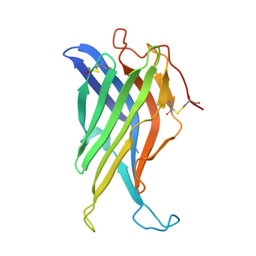
Crystal Structure of the Conserved Domain of the Dc Lysosomal Associated Membrane Protein: Implications for the Lysosomal Glycocalyx.

Wilke, S.Krausze, J.Bussow, K.

(2012) BMC Biol 10: 62

  • DOI: https://doi.org/10.1186/1741-7007-10-62
  • Primary Citation of Related Structures:  
    4AKM

  • PubMed Abstract: 

    The family of lysosome-associated membrane proteins (LAMP) comprises the multifunctional, ubiquitous LAMP-1 and LAMP-2, and the cell type-specific proteins DC-LAMP (LAMP-3), BAD-LAMP (UNC-46, C20orf103) and macrosialin (CD68). LAMPs have been implicated in a multitude of cellular processes, including phagocytosis, autophagy, lipid transport and aging. LAMP-2 isoform A acts as a receptor in chaperone-mediated autophagy. LAMP-2 deficiency causes the fatal Danon disease. The abundant proteins LAMP-1 and LAMP-2 are major constituents of the glycoconjugate coat present on the inside of the lysosomal membrane, the 'lysosomal glycocalyx'. The LAMP family is characterized by a conserved domain of 150 to 200 amino acids with two disulfide bonds.


  • Organizational Affiliation

    Department of Molecular Structural Biology, Helmholtz Centre for Infection Research, Inhoffenstr, 7, 38124 Braunschweig, Germany.


Macromolecules
Find similar proteins by:  (by identity cutoff)  |  3D Structure
Entity ID: 1
MoleculeChains Sequence LengthOrganismDetailsImage
LYSOSOME-ASSOCIATED MEMBRANE GLYCOPROTEIN 3
A, B
166Homo sapiensMutation(s): 1 
UniProt & NIH Common Fund Data Resources
Find proteins for Q9UQV4 (Homo sapiens)
Explore Q9UQV4 
Go to UniProtKB:  Q9UQV4
PHAROS:  Q9UQV4
GTEx:  ENSG00000078081 
Entity Groups  
Sequence Clusters30% Identity50% Identity70% Identity90% Identity95% Identity100% Identity
UniProt GroupQ9UQV4
Sequence Annotations
Expand
  • Reference Sequence
Experimental Data & Validation

Experimental Data

  • Method: X-RAY DIFFRACTION
  • Resolution: 2.69 Å
  • R-Value Free: 0.249 
  • R-Value Work: 0.227 
  • R-Value Observed: 0.228 
  • Space Group: P 31
Unit Cell:
Length ( Å )Angle ( ˚ )
a = 52.6α = 90
b = 52.6β = 90
c = 141.98γ = 120
Software Package:
Software NamePurpose
PHENIXrefinement
XDSdata reduction
XSCALEdata scaling
PHENIXphasing

Structure Validation

View Full Validation Report


Ligand Structure Quality Assessment 


Entry History 

Deposition Data

Revision History  (Full details and data files)

  • Version 1.0: 2012-08-15
    Type: Initial release
  • Version 1.1: 2017-07-12
    Changes: Refinement description
  • Version 1.2: 2019-07-17
    Changes: Data collection, Derived calculations
  • Version 1.3: 2020-07-29
    Type: Remediation
    Reason: Carbohydrate remediation
    Changes: Data collection, Derived calculations, Other, Structure summary