3ZS5

Structural basis for kinase selectivity of three clinical p38alpha inhibitors


Experimental Data Snapshot

  • Method: X-RAY DIFFRACTION
  • Resolution: 1.60 Å
  • R-Value Free: 0.215 
  • R-Value Work: 0.184 
  • R-Value Observed: 0.186 

wwPDB Validation   3D Report Full Report


Ligand Structure Quality Assessment 


This is version 1.5 of the entry. See complete history


Literature

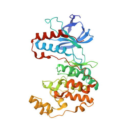
X-ray structure of p38 alpha bound to TAK-715: comparison with three classic inhibitors.

Azevedo, R.van Zeeland, M.Raaijmakers, H.Kazemier, B.de Vlieg, J.Oubrie, A.

(2012) Acta Crystallogr D Biol Crystallogr 68: 1041-1050

  • DOI: https://doi.org/10.1107/S090744491201997X
  • Primary Citation of Related Structures:  
    3ZS5, 3ZSG, 3ZSH, 3ZSI

  • PubMed Abstract: 

    The p38α mitogen-activated protein kinase regulates the synthesis of pro-inflammatory cytokines in response to stimulation by a diverse set of stress signals. Various different chemotypes and clinical candidates that inhibit p38α function have been reported over the years. In this publication, the novel structure of p38α cocrystallized with the clinical candidate TAK-715 is reported. Owing to the impact of crystallization conditions on the conformation of protein kinases (and in particular p38α), the structures of complexes of p38α with SB-203580, SCIO-469 and VX-745 have also been determined to enable in-depth comparison of ligand-induced protein conformations. The impact of experimental conditions on p38α-inhibitor complex structures, most importantly soaking versus cocrystallization, is discussed. Analysis of the structures and quantification of the protein-ligand interactions couples ligand-induced protein conformations to the number of interactions and to inhibitor selectivity against the human kinome. This shows that for the design of novel kinase inhibitors, selectivity is best obtained through maximization of the number of interactions throughout the ATP pocket and the exploitation of specific features in the active site.


  • Organizational Affiliation

    Merck Research Laboratories, MSD, PO Box 20, 5340 BH Oss, The Netherlands.


Macromolecules
Find similar proteins by:  (by identity cutoff)  |  3D Structure
Entity ID: 1
MoleculeChains Sequence LengthOrganismDetailsImage
MITOGEN-ACTIVATED PROTEIN KINASE 14362Homo sapiensMutation(s): 0 
EC: 2.7.11.24
UniProt & NIH Common Fund Data Resources
Find proteins for Q16539 (Homo sapiens)
Explore Q16539 
Go to UniProtKB:  Q16539
PHAROS:  Q16539
GTEx:  ENSG00000112062 
Entity Groups  
Sequence Clusters30% Identity50% Identity70% Identity90% Identity95% Identity100% Identity
UniProt GroupQ16539
Sequence Annotations
Expand
  • Reference Sequence
Experimental Data & Validation

Experimental Data

  • Method: X-RAY DIFFRACTION
  • Resolution: 1.60 Å
  • R-Value Free: 0.215 
  • R-Value Work: 0.184 
  • R-Value Observed: 0.186 
  • Space Group: P 21 21 21
Unit Cell:
Length ( Å )Angle ( ˚ )
a = 67.492α = 90
b = 70.036β = 90
c = 75.01γ = 90
Software Package:
Software NamePurpose
REFMACrefinement
MOSFLMdata reduction
SCALAdata scaling
REFMACphasing

Structure Validation

View Full Validation Report


Ligand Structure Quality Assessment 


Entry History 

Deposition Data

Revision History  (Full details and data files)

  • Version 1.0: 2012-07-04
    Type: Initial release
  • Version 1.1: 2012-07-25
    Changes: Database references
  • Version 1.2: 2012-08-15
    Changes: Database references
  • Version 1.3: 2018-02-07
    Changes: Database references, Structure summary
  • Version 1.4: 2019-05-08
    Changes: Data collection, Experimental preparation
  • Version 1.5: 2020-07-29
    Type: Remediation
    Reason: Carbohydrate remediation
    Changes: Data collection, Derived calculations, Other, Structure summary