3ZH9

Bacillus subtilis DNA clamp loader delta protein (YqeN)


Experimental Data Snapshot

  • Method: X-RAY DIFFRACTION
  • Resolution: 2.10 Å
  • R-Value Free: 0.239 
  • R-Value Work: 0.192 
  • R-Value Observed: 0.194 

wwPDB Validation   3D Report Full Report


This is version 1.1 of the entry. See complete history


Literature

Insights Into the Structure and Assembly of the Bacillus Subtilis Clamp-Loader Complex and its Interaction with the Replicative Helicase.

Afonso, J.P.Chintakayala, K.Suwannachart, C.Sedelnikova, S.Giles, K.Hoyes, J.B.Soultanas, P.Rafferty, J.B.Oldham, N.J.

(2013) Nucleic Acids Res 41: 5115

  • DOI: https://doi.org/10.1093/nar/gkt173
  • Primary Citation of Related Structures:  
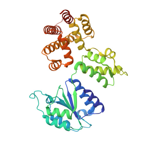
    3ZH9

  • PubMed Abstract: 

    The clamp-loader complex plays a crucial role in DNA replication by loading the β-clamp onto primed DNA to be used by the replicative polymerase. Relatively little is known about the stoichiometry, structure and assembly pathway of this complex, and how it interacts with the replicative helicase, in Gram-positive organisms. Analysis of full and partial complexes by mass spectrometry revealed that a hetero-pentameric τ3-δ-δ' Bacillus subtilis clamp-loader assembles via multiple pathways, which differ from those exhibited by the Gram-negative model Escherichia coli. Based on this information, a homology model of the B. subtilis τ3-δ-δ' complex was constructed, which revealed the spatial positioning of the full C-terminal τ domain. The structure of the δ subunit was determined by X-ray crystallography and shown to differ from that of E. coli in the nature of the amino acids comprising the τ and δ' binding regions. Most notably, the τ-δ interaction appears to be hydrophilic in nature compared with the hydrophobic interaction in E. coli. Finally, the interaction between τ3 and the replicative helicase DnaB was driven by ATP/Mg(2+) conformational changes in DnaB, and evidence is provided that hydrolysis of one ATP molecule by the DnaB hexamer is sufficient to stabilize its interaction with τ3.


  • Organizational Affiliation

    School of Chemistry, University of Nottingham, University Park, Nottingham NG7 2RD, UK.


Macromolecules
Find similar proteins by:  (by identity cutoff)  |  3D Structure
Entity ID: 1
MoleculeChains Sequence LengthOrganismDetailsImage
DELTAA [auth B]347Bacillus subtilis subsp. subtilis str. 168Mutation(s): 0 
Gene Names: YQEN
EC: 2.7.7.7
UniProt
Find proteins for P54459 (Bacillus subtilis (strain 168))
Explore P54459 
Go to UniProtKB:  P54459
Entity Groups  
Sequence Clusters30% Identity50% Identity70% Identity90% Identity95% Identity100% Identity
UniProt GroupP54459
Sequence Annotations
Expand
  • Reference Sequence
Small Molecules
Ligands 2 Unique
IDChains Name / Formula / InChI Key2D Diagram3D Interactions
SO4
Query on SO4

Download Ideal Coordinates CCD File 
B
C [auth B]
E [auth B]
G [auth B]
H [auth B]
B,
C [auth B],
E [auth B],
G [auth B],
H [auth B],
J [auth B],
M [auth B]
SULFATE ION
O4 S
QAOWNCQODCNURD-UHFFFAOYSA-L
GOL
Query on GOL

Download Ideal Coordinates CCD File 
D [auth B]
F [auth B]
I [auth B]
K [auth B]
L [auth B]
D [auth B],
F [auth B],
I [auth B],
K [auth B],
L [auth B],
N [auth B],
O [auth B]
GLYCEROL
C3 H8 O3
PEDCQBHIVMGVHV-UHFFFAOYSA-N
Experimental Data & Validation

Experimental Data

  • Method: X-RAY DIFFRACTION
  • Resolution: 2.10 Å
  • R-Value Free: 0.239 
  • R-Value Work: 0.192 
  • R-Value Observed: 0.194 
  • Space Group: P 43 21 2
Unit Cell:
Length ( Å )Angle ( ˚ )
a = 81.6α = 90
b = 81.6β = 90
c = 157.67γ = 90
Software Package:
Software NamePurpose
PHENIXrefinement
iMOSFLMdata reduction
SCALAdata scaling
SOLVEphasing

Structure Validation

View Full Validation Report


Entry History 

Deposition Data

Revision History  (Full details and data files)

  • Version 1.0: 2013-04-03
    Type: Initial release
  • Version 1.1: 2013-05-22
    Changes: Database references