3WXA

X-ray crystal structural analysis of the complex between ALG-2 and Sec31A peptide


Experimental Data Snapshot

  • Method: X-RAY DIFFRACTION
  • Resolution: 2.36 Å
  • R-Value Free: 0.263 
  • R-Value Work: 0.225 
  • R-Value Observed: 0.230 

wwPDB Validation   3D Report Full Report


This is version 1.2 of the entry. See complete history


Literature

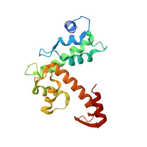
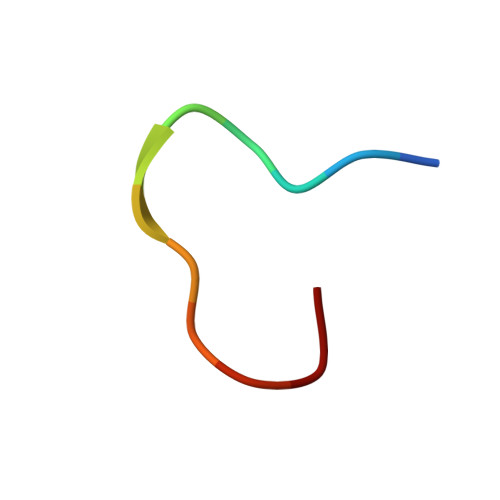
Structural Analysis of the Complex between Penta-EF-Hand ALG-2 Protein and Sec31A Peptide Reveals a Novel Target Recognition Mechanism of ALG-2

Takahashi, T.Kojima, K.Zhang, W.Sasaki, K.Ito, M.Suzuki, H.Kawasaki, M.Wakatsuki, S.Takahara, T.Shibata, H.Maki, M.

(2015) Int J Mol Sci 16: 3677-3699

  • DOI: https://doi.org/10.3390/ijms16023677
  • Primary Citation of Related Structures:  
    3WXA

  • PubMed Abstract: 

    ALG-2, a 22-kDa penta-EF-hand protein, is involved in cell death, signal transduction, membrane trafficking, etc., by interacting with various proteins in mammalian cells in a Ca2+-dependent manner. Most known ALG-2-interacting proteins contain proline-rich regions in which either PPYPXnYP (type 1 motif) or PXPGF (type 2 motif) is commonly found. Previous X-ray crystal structural analysis of the complex between ALG-2 and an ALIX peptide revealed that the peptide binds to the two hydrophobic pockets. In the present study, we resolved the crystal structure of the complex between ALG-2 and a peptide of Sec31A (outer shell component of coat complex II, COPII; containing the type 2 motif) and found that the peptide binds to the third hydrophobic pocket (Pocket 3). While amino acid substitution of Phe85, a Pocket 3 residue, with Ala abrogated the interaction with Sec31A, it did not affect the interaction with ALIX. On the other hand, amino acid substitution of Tyr180, a Pocket 1 residue, with Ala caused loss of binding to ALIX, but maintained binding to Sec31A. We conclude that ALG-2 recognizes two types of motifs at different hydrophobic surfaces. Furthermore, based on the results of serial mutational analysis of the ALG-2-binding sites in Sec31A, the type 2 motif was newly defined.


  • Organizational Affiliation

    Department of Applied Molecular Biosciences, Graduate School of Bioagricultural Sciences, Nagoya University, Furo-cho, Chikusa-ku, Nagoya 464-8601, Japan. takahashi.takeshi@a.mbox.nagoya-u.ac.jp.


Macromolecules
Find similar proteins by:  (by identity cutoff)  |  3D Structure
Entity ID: 1
MoleculeChains Sequence LengthOrganismDetailsImage
Programmed cell death protein 6
A, B
172Homo sapiensMutation(s): 0 
Gene Names: PDCD6ALG2
UniProt & NIH Common Fund Data Resources
Find proteins for O75340 (Homo sapiens)
Explore O75340 
Go to UniProtKB:  O75340
PHAROS:  O75340
GTEx:  ENSG00000249915 
Entity Groups  
Sequence Clusters30% Identity50% Identity70% Identity90% Identity95% Identity100% Identity
UniProt GroupO75340
Sequence Annotations
Expand
  • Reference Sequence

Find similar proteins by:  Sequence   |   3D Structure  

Entity ID: 2
MoleculeChains Sequence LengthOrganismDetailsImage
Protein transport protein Sec31A
C, D
12Homo sapiensMutation(s): 0 
UniProt & NIH Common Fund Data Resources
Find proteins for O94979 (Homo sapiens)
Explore O94979 
Go to UniProtKB:  O94979
PHAROS:  O94979
GTEx:  ENSG00000138674 
Entity Groups  
Sequence Clusters30% Identity50% Identity70% Identity90% Identity95% Identity100% Identity
UniProt GroupO94979
Sequence Annotations
Expand
  • Reference Sequence
Experimental Data & Validation

Experimental Data

  • Method: X-RAY DIFFRACTION
  • Resolution: 2.36 Å
  • R-Value Free: 0.263 
  • R-Value Work: 0.225 
  • R-Value Observed: 0.230 
  • Space Group: P 64
Unit Cell:
Length ( Å )Angle ( ˚ )
a = 81.973α = 90
b = 81.973β = 90
c = 103.232γ = 120
Software Package:
Software NamePurpose
SERGUIdata collection
MOLREPphasing
PHENIXrefinement
HKL-2000data reduction
HKL-2000data scaling

Structure Validation

View Full Validation Report


Entry History 

Deposition Data

Revision History  (Full details and data files)

  • Version 1.0: 2015-03-11
    Type: Initial release
  • Version 1.1: 2017-11-22
    Changes: Refinement description
  • Version 1.2: 2023-11-08
    Changes: Data collection, Database references, Derived calculations, Refinement description