3VQH

Bromine SAD partially resolves multiple binding modes for PKA inhibitor H-89


Experimental Data Snapshot

  • Method: X-RAY DIFFRACTION
  • Resolution: 1.95 Å
  • R-Value Free: 0.234 
  • R-Value Work: 0.190 
  • R-Value Observed: 0.192 

wwPDB Validation   3D Report Full Report


Ligand Structure Quality Assessment 


This is version 1.2 of the entry. See complete history


Literature

Anomalous dispersion analysis of inhibitor flexibility: a case study of the kinase inhibitor H-89

Pflug, A.Johnson, K.A.Engh, R.A.

(2012) Acta Crystallogr Sect F Struct Biol Cryst Commun 68: 873-877

  • DOI: https://doi.org/10.1107/S1744309112028655
  • Primary Citation of Related Structures:  
    3VQH

  • PubMed Abstract: 

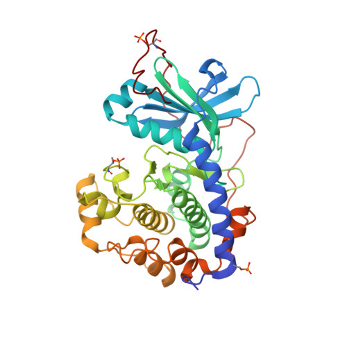
    With its ability to show the interactions between drug-target proteins and small-molecule ligands, X-ray crystallography is an essential tool in drug-discovery programmes. However, its usefulness can be limited by crystallization artifacts or by the data resolution, and in particular when assumptions of unimodal binding (and isotropic motion) do not apply. Discrepancies between the modelled crystal structure and the physiological range of structures generally prevent quantitative estimation of binding energies. Improved crystal structure resolution will often not aid energy estimation because the conditions which provide the highest rigidity and resolution are not likely to reflect physiological conditions. Instead, strategies must be employed to measure and model flexibility and multiple binding modes to supplement crystallographic information. One useful tool is the use of anomalous dispersion for small molecules that contain suitable atoms. Here, an analysis of the binding of the kinase inhibitor H-89 to protein kinase A (PKA) is presented. H-89 contains a bromobenzene moiety that apparently binds with multiple conformations in the kinase ATP pocket. Using anomalous dispersion methods, it was possible to resolve these conformations into two distinct binding geometries.


  • Organizational Affiliation

    Norwegian Structural Biology Centre, Department of Chemistry, University of Tromsø, N-9037 Tromsø, Norway.


Macromolecules
Find similar proteins by:  (by identity cutoff)  |  3D Structure
Entity ID: 1
MoleculeChains Sequence LengthOrganismDetailsImage
cAMP-dependent protein kinase catalytic subunit alpha351Homo sapiensMutation(s): 0 
Gene Names: PKACAPRKACA
EC: 2.7.11.11
UniProt & NIH Common Fund Data Resources
Find proteins for P17612 (Homo sapiens)
Explore P17612 
Go to UniProtKB:  P17612
PHAROS:  P17612
GTEx:  ENSG00000072062 
Entity Groups  
Sequence Clusters30% Identity50% Identity70% Identity90% Identity95% Identity100% Identity
UniProt GroupP17612
Sequence Annotations
Expand
  • Reference Sequence

Find similar proteins by:  Sequence   |   3D Structure  

Entity ID: 2
MoleculeChains Sequence LengthOrganismDetailsImage
cAMP-dependent protein kinase inhibitor alpha20Homo sapiensMutation(s): 0 
UniProt & NIH Common Fund Data Resources
Find proteins for P61925 (Homo sapiens)
Explore P61925 
Go to UniProtKB:  P61925
PHAROS:  P61925
GTEx:  ENSG00000171033 
Entity Groups  
Sequence Clusters30% Identity50% Identity70% Identity90% Identity95% Identity100% Identity
UniProt GroupP61925
Sequence Annotations
Expand
  • Reference Sequence
Small Molecules
Ligands 2 Unique
IDChains Name / Formula / InChI Key2D Diagram3D Interactions
IQB
Query on IQB

Download Ideal Coordinates CCD File 
C [auth A]N-[2-(4-BROMOCINNAMYLAMINO)ETHYL]-5-ISOQUINOLINE SULFONAMIDE
C20 H20 Br N3 O2 S
ZKZXNDJNWUTGDK-NSCUHMNNSA-N
MPD
Query on MPD

Download Ideal Coordinates CCD File 
D [auth A](4S)-2-METHYL-2,4-PENTANEDIOL
C6 H14 O2
SVTBMSDMJJWYQN-YFKPBYRVSA-N
Modified Residues  2 Unique
IDChains TypeFormula2D DiagramParent
SEP
Query on SEP
A
L-PEPTIDE LINKINGC3 H8 N O6 PSER
TPO
Query on TPO
A
L-PEPTIDE LINKINGC4 H10 N O6 PTHR
Binding Affinity Annotations 
IDSourceBinding Affinity
IQB BindingDB:  3VQH Ki: min: 48, max: 50 (nM) from 2 assay(s)
Kd: 23 (nM) from 1 assay(s)
IC50: min: 73, max: 100 (nM) from 2 assay(s)
PDBBind:  3VQH IC50: 1200 (nM) from 1 assay(s)
Experimental Data & Validation

Experimental Data

  • Method: X-RAY DIFFRACTION
  • Resolution: 1.95 Å
  • R-Value Free: 0.234 
  • R-Value Work: 0.190 
  • R-Value Observed: 0.192 
  • Space Group: P 21 21 21
Unit Cell:
Length ( Å )Angle ( ˚ )
a = 72.73α = 90
b = 75.18β = 90
c = 80.33γ = 90
Software Package:
Software NamePurpose
MOLREPphasing
REFMACrefinement
XDSdata reduction
XSCALEdata scaling

Structure Validation

View Full Validation Report


Ligand Structure Quality Assessment 


Entry History 

Deposition Data

Revision History  (Full details and data files)

  • Version 1.0: 2012-08-15
    Type: Initial release
  • Version 1.1: 2014-04-30
    Changes: Database references
  • Version 1.2: 2023-11-08
    Changes: Data collection, Database references, Derived calculations, Refinement description