3UVV

Crystal Structure of the ligand binding domains of the thyroid receptor:retinoid X receptor complexed with 3,3',5 triiodo-L-thyronine and 9-cis retinoic acid


Experimental Data Snapshot

  • Method: X-RAY DIFFRACTION
  • Resolution: 2.95 Å
  • R-Value Free: 0.250 
  • R-Value Work: 0.201 
  • R-Value Observed: 0.203 

wwPDB Validation   3D Report Full Report


Ligand Structure Quality Assessment 


This is version 2.0 of the entry. See complete history


Literature

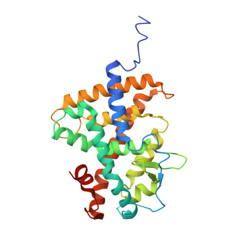
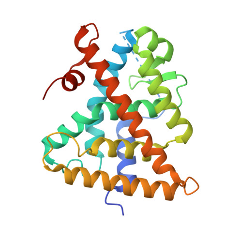
Structural basis for negative cooperativity within agonist-bound TR:RXR heterodimers.

Putcha, B.D.Wright, E.Brunzelle, J.S.Fernandez, E.J.

(2012) Proc Natl Acad Sci U S A 109: 6084-6087

  • DOI: https://doi.org/10.1073/pnas.1119852109
  • Primary Citation of Related Structures:  
    3UVV

  • PubMed Abstract: 

    Thyroid hormones such as 3,3',5 triiodo-L-thyronine (T3) control numerous aspects of mammalian development and metabolism. The actions of such hormones are mediated by specific thyroid hormone receptors (TRs). TR belongs to the nuclear receptor family of modular transcription factors that binds to specific DNA-response elements within target promoters. These receptors can function as homo- or heterodimers such as TR:9-cis retinoic acid receptor (RXR). Here, we present the atomic resolution structure of the TRα•T3:RXRα•9-cis retinoic acid (9c) ligand binding domain heterodimer complex at 2.95 Å along with T3 hormone binding and dissociation and coactivator binding studies. Our data provide a structural basis for allosteric communication between T3 and 9c and negative cooperativity between their binding pockets. In this structure, both TR and RXR are in the active state conformation for optimal binding to coactivator proteins. However, the structure of TR•T3 within TR•T3:RXR•9c is in a relative state of disorder, and the observed kinetics of binding show that T3 dissociates more rapidly from TR•T3:RXR•9c than from TR•T3:RXR. Also, coactivator binding studies with a steroid receptor coactivator-1 (receptor interaction domains 1-3) fragment show lower affinities (K(a)) for TR•T3:RXR•9c than TR•T3:RXR. Our study corroborates previously reported observations from cell-based and binding studies and offers a structural mechanism for the repression of TR•T3:RXR transactivation by RXR agonists. Furthermore, the recent discoveries of multiple endogenous RXR agonists that mediate physiological tasks such as lipid biosynthesis underscore the pharmacological importance of negative cooperativity in ligand binding within TR:RXR heterodimers.


  • Organizational Affiliation

    University of Tennessee, Knoxville, TN 37996, USA.


Macromolecules
Find similar proteins by:  (by identity cutoff)  |  3D Structure
Entity ID: 1
MoleculeChains Sequence LengthOrganismDetailsImage
Thyroid hormone receptor alpha265Gallus gallusMutation(s): 0 
Gene Names: THRANR1A1
UniProt
Find proteins for P04625 (Gallus gallus)
Explore P04625 
Go to UniProtKB:  P04625
Entity Groups  
Sequence Clusters30% Identity50% Identity70% Identity90% Identity95% Identity100% Identity
UniProt GroupP04625
Sequence Annotations
Expand
  • Reference Sequence
Find similar proteins by:  (by identity cutoff)  |  3D Structure
Entity ID: 2
MoleculeChains Sequence LengthOrganismDetailsImage
Retinoic acid receptor RXR-alpha244Homo sapiensMutation(s): 0 
Gene Names: RXRANR2B1
UniProt & NIH Common Fund Data Resources
Find proteins for P19793 (Homo sapiens)
Explore P19793 
Go to UniProtKB:  P19793
PHAROS:  P19793
GTEx:  ENSG00000186350 
Entity Groups  
Sequence Clusters30% Identity50% Identity70% Identity90% Identity95% Identity100% Identity
UniProt GroupP19793
Sequence Annotations
Expand
  • Reference Sequence
Small Molecules
Ligands 2 Unique
IDChains Name / Formula / InChI Key2D Diagram3D Interactions
T3
Query on T3

Download Ideal Coordinates CCD File 
C [auth A]3,5,3'TRIIODOTHYRONINE
C15 H12 I3 N O4
AUYYCJSJGJYCDS-LBPRGKRZSA-N
9CR
Query on 9CR

Download Ideal Coordinates CCD File 
D [auth B](9cis)-retinoic acid
C20 H28 O2
SHGAZHPCJJPHSC-ZVCIMWCZSA-N
Binding Affinity Annotations 
IDSourceBinding Affinity
T3 BindingDB:  3UVV Ki: min: 0.08, max: 2.33 (nM) from 9 assay(s)
Kd: min: 0.06, max: 0.17 (nM) from 8 assay(s)
IC50: min: 0.24, max: 500 (nM) from 7 assay(s)
EC50: min: 0.41, max: 15 (nM) from 13 assay(s)
9CR BindingDB:  3UVV Ki: min: 3.8, max: 583 (nM) from 12 assay(s)
Kd: min: 1.5, max: 1810 (nM) from 20 assay(s)
IC50: min: 4, max: 82 (nM) from 5 assay(s)
EC50: min: 1.5, max: 316 (nM) from 22 assay(s)
Experimental Data & Validation

Experimental Data

  • Method: X-RAY DIFFRACTION
  • Resolution: 2.95 Å
  • R-Value Free: 0.250 
  • R-Value Work: 0.201 
  • R-Value Observed: 0.203 
  • Space Group: C 2 2 21
Unit Cell:
Length ( Å )Angle ( ˚ )
a = 129.13α = 90
b = 165.32β = 90
c = 85.1γ = 90
Software Package:
Software NamePurpose
AMoREphasing
BUSTERrefinement
HKL-2000data reduction
HKL-2000data scaling

Structure Validation

View Full Validation Report


Ligand Structure Quality Assessment 


Entry History 

Deposition Data

Revision History  (Full details and data files)

  • Version 1.0: 2012-04-18
    Type: Initial release
  • Version 1.1: 2012-05-02
    Changes: Database references
  • Version 1.2: 2016-11-16
    Changes: Non-polymer description
  • Version 1.3: 2023-09-13
    Changes: Data collection, Database references, Derived calculations, Refinement description
  • Version 2.0: 2023-11-15
    Changes: Atomic model, Data collection