3UMK

X-ray structure of the E2 domain of the human amyloid precursor protein (APP) in complex with copper


Experimental Data Snapshot

  • Method: X-RAY DIFFRACTION
  • Resolution: 2.60 Å
  • R-Value Free: 0.239 
  • R-Value Work: 0.206 
  • R-Value Observed: 0.206 

wwPDB Validation   3D Report Full Report


This is version 1.3 of the entry. See complete history


Literature

Metal Binding Dictates Conformation and Function of the Amyloid Precursor Protein (APP) E2 Domain.

Dahms, S.O.Konnig, I.Roeser, D.Guhrs, K.H.Mayer, M.C.Kaden, D.Multhaup, G.Than, M.E.

(2012) J Mol Biol 416: 438-452

  • DOI: https://doi.org/10.1016/j.jmb.2011.12.057
  • Primary Citation of Related Structures:  
    3UMH, 3UMI, 3UMK

  • PubMed Abstract: 

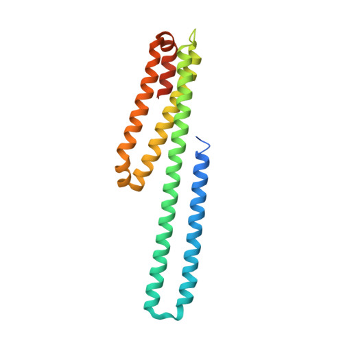
    The amyloid precursor protein (APP) and its neurotoxic cleavage product Aβ are key players in the development of Alzheimer's disease and appear essential for neuronal development and cell homeostasis in mammals. Proteolytic processing of APP is influenced by metal ions, protein ligands and its oligomerization state. However, the structural basis and functional mechanism of APP regulation are hitherto largely unknown. Here we identified a metal-dependent molecular switch located within the E2 domain of APP containing four evolutionary highly conserved histidine residues. Three X-ray structures of the metal-bound molecule were solved at 2.6-2.0 Å resolution. Using protein crystallographic and biochemical methods, we characterized this novel high-affinity binding site within the E2 domain that binds competitively to copper and zinc at physiological concentrations. Metal-specific coordination spheres induce large conformational changes and enforce distinct structural states, most likely regulating the physiological function of APP and its processing in Alzheimer's disease.


  • Organizational Affiliation

    Protein Crystallography Group, Leibniz Institute for Age Research - Fritz Lipmann Institute (FLI), Beutenbergstr. 11, 07745 Jena, Germany.


Macromolecules
Find similar proteins by:  (by identity cutoff)  |  3D Structure
Entity ID: 1
MoleculeChains Sequence LengthOrganismDetailsImage
Amyloid beta A4 protein211Homo sapiensMutation(s): 0 
UniProt & NIH Common Fund Data Resources
Find proteins for P05067 (Homo sapiens)
Explore P05067 
Go to UniProtKB:  P05067
PHAROS:  P05067
GTEx:  ENSG00000142192 
Entity Groups  
Sequence Clusters30% Identity50% Identity70% Identity90% Identity95% Identity100% Identity
UniProt GroupP05067
Sequence Annotations
Expand
  • Reference Sequence
Small Molecules
Ligands 3 Unique
IDChains Name / Formula / InChI Key2D Diagram3D Interactions
CD
Query on CD

Download Ideal Coordinates CCD File 
D [auth A]
E [auth A]
F [auth A]
H [auth A]
I [auth A]
D [auth A],
E [auth A],
F [auth A],
H [auth A],
I [auth A],
J [auth A],
K [auth A]
CADMIUM ION
Cd
WLZRMCYVCSSEQC-UHFFFAOYSA-N
CU
Query on CU

Download Ideal Coordinates CCD File 
C [auth A],
G [auth A]
COPPER (II) ION
Cu
JPVYNHNXODAKFH-UHFFFAOYSA-N
ACT
Query on ACT

Download Ideal Coordinates CCD File 
B [auth A]ACETATE ION
C2 H3 O2
QTBSBXVTEAMEQO-UHFFFAOYSA-M
Experimental Data & Validation

Experimental Data

  • Method: X-RAY DIFFRACTION
  • Resolution: 2.60 Å
  • R-Value Free: 0.239 
  • R-Value Work: 0.206 
  • R-Value Observed: 0.206 
  • Space Group: P 43
Unit Cell:
Length ( Å )Angle ( ˚ )
a = 39.76α = 90
b = 39.76β = 90
c = 126.42γ = 90
Software Package:
Software NamePurpose
SHARPphasing
CNSrefinement
MOSFLMdata reduction
SCALAdata scaling

Structure Validation

View Full Validation Report


Entry History 

Deposition Data

Revision History  (Full details and data files)

  • Version 1.0: 2012-01-25
    Type: Initial release
  • Version 1.1: 2012-02-22
    Changes: Database references
  • Version 1.2: 2017-11-08
    Changes: Advisory, Refinement description
  • Version 1.3: 2024-02-28
    Changes: Advisory, Data collection, Database references, Derived calculations