X-ray Structures of Magnesium and Manganese Complexes with the N-Terminal Domain of Calmodulin: Insights into the Mechanism and Specificity of Metal Ion Binding to an EF-Hand.
Senguen, F.T., Grabarek, Z.(2012) Biochemistry 51: 6182-6194
- PubMed: 22803592
- DOI: https://doi.org/10.1021/bi300698h
- Primary Citation of Related Structures:
3UCT, 3UCW, 3UCY - PubMed Abstract:
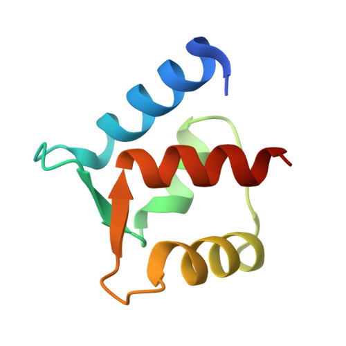
Calmodulin (CaM), a member of the EF-hand superfamily, regulates many aspects of cell function by responding specifically to micromolar concentrations of Ca(2+) in the presence of an ~1000-fold higher concentration of cellular Mg(2+). To explain the structural basis of metal ion binding specificity, we have determined the X-ray structures of the N-terminal domain of calmodulin (N-CaM) in complexes with Mg(2+), Mn(2+), and Zn(2+). In contrast to Ca(2+), which induces domain opening in CaM, octahedrally coordinated Mg(2+) and Mn(2+) stabilize the closed-domain, apo-like conformation, while tetrahedrally coordinated Zn(2+) ions bind at the protein surface and do not compete with Ca(2+). The relative positions of bound Mg(2+) and Mn(2+) within the EF-hand loops are similar to those of Ca(2+); however, the Glu side chain at position 12 of the loop, whose bidentate interaction with Ca(2+) is critical for domain opening, does not bind directly to either Mn(2+) or Mg(2+), and the vacant ligand position is occupied by a water molecule. We conclude that this critical interaction is prevented by specific stereochemical constraints imposed on the ligands by the EF-hand β-scaffold. The structures suggest that Mg(2+) contributes to the switching off of calmodulin activity and possibly other EF-hand proteins at the resting levels of Ca(2+). The Mg(2+)-bound N-CaM structure also provides a unique view of a transiently bound hydrated metal ion and suggests a role for the hydration water in the metal-induced conformational change.
Organizational Affiliation:
Boston Biomedical Research Institute, Watertown, MA 02472, USA.