3U5V

Crystal structure of Max-E47


Experimental Data Snapshot

  • Method: X-RAY DIFFRACTION
  • Resolution: 1.70 Å
  • R-Value Free: 0.235 
  • R-Value Work: 0.207 
  • R-Value Observed: 0.208 

wwPDB Validation   3D Report Full Report


This is version 1.2 of the entry. See complete history


Literature

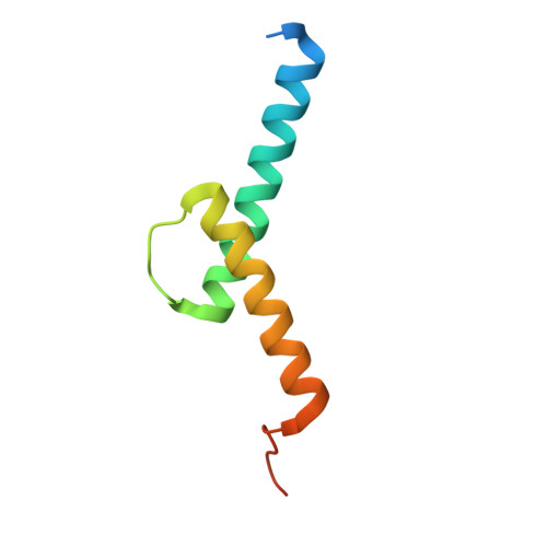
Crystal structure of the minimalist max-e47 protein chimera.

Ahmadpour, F.Ghirlando, R.De Jong, A.T.Gloyd, M.Shin, J.A.Guarne, A.

(2012) PLoS One 7: e32136-e32136

  • DOI: https://doi.org/10.1371/journal.pone.0032136
  • Primary Citation of Related Structures:  
    3U5V

  • PubMed Abstract: 

    Max-E47 is a protein chimera generated from the fusion of the DNA-binding basic region of Max and the dimerization region of E47, both members of the basic region/helix-loop-helix (bHLH) superfamily of transcription factors. Like native Max, Max-E47 binds with high affinity and specificity to the E-box site, 5'-CACGTG, both in vivo and in vitro. We have determined the crystal structure of Max-E47 at 1.7 Å resolution, and found that it associates to form a well-structured dimer even in the absence of its cognate DNA. Analytical ultracentrifugation confirms that Max-E47 is dimeric even at low micromolar concentrations, indicating that the Max-E47 dimer is stable in the absence of DNA. Circular dichroism analysis demonstrates that both non-specific DNA and the E-box site induce similar levels of helical secondary structure in Max-E47. These results suggest that Max-E47 may bind to the E-box following the two-step mechanism proposed for other bHLH proteins. In this mechanism, a rapid step where protein binds to DNA without sequence specificity is followed by a slow step where specific protein:DNA interactions are fine-tuned, leading to sequence-specific recognition. Collectively, these results show that the designed Max-E47 protein chimera behaves both structurally and functionally like its native counterparts.


  • Organizational Affiliation

    Department of Biochemistry and Biomedical Sciences, McMaster University, Hamilton, Ontario, Canada.


Macromolecules
Find similar proteins by:  (by identity cutoff)  |  3D Structure
Entity ID: 1
MoleculeChains Sequence LengthOrganismDetailsImage
Protein max, Transcription factor E2-alpha chimera76Mus musculusHomo sapiens
This entity is chimeric
Mutation(s): 0 
Gene Names: MaxMynBHLHB21E2AITF1TCF3
UniProt & NIH Common Fund Data Resources
Find proteins for P28574 (Mus musculus)
Explore P28574 
Go to UniProtKB:  P28574
Find proteins for P15923 (Homo sapiens)
Explore P15923 
Go to UniProtKB:  P15923
PHAROS:  P15923
GTEx:  ENSG00000071564 
Entity Groups  
Sequence Clusters30% Identity50% Identity70% Identity90% Identity95% Identity100% Identity
UniProt GroupsP15923P28574
Sequence Annotations
Expand
  • Reference Sequence
Experimental Data & Validation

Experimental Data

  • Method: X-RAY DIFFRACTION
  • Resolution: 1.70 Å
  • R-Value Free: 0.235 
  • R-Value Work: 0.207 
  • R-Value Observed: 0.208 
  • Space Group: I 21 21 21
Unit Cell:
Length ( Å )Angle ( ˚ )
a = 37.48α = 90
b = 49.466β = 90
c = 74.1γ = 90
Software Package:
Software NamePurpose
CBASSdata collection
PHASESphasing
PHENIXrefinement
HKL-2000data reduction
HKL-2000data scaling

Structure Validation

View Full Validation Report


Entry History 

Deposition Data

Revision History  (Full details and data files)

  • Version 1.0: 2012-03-21
    Type: Initial release
  • Version 1.1: 2017-07-26
    Changes: Advisory, Refinement description, Source and taxonomy
  • Version 1.2: 2023-09-13
    Changes: Advisory, Data collection, Database references, Derived calculations, Refinement description