3TS8

Crystal structure of a multidomain human p53 tetramer bound to the natural CDKN1A(p21) p53-response element


Experimental Data Snapshot

  • Method: X-RAY DIFFRACTION
  • Resolution: 2.80 Å
  • R-Value Free: 0.283 
  • R-Value Work: 0.236 
  • R-Value Observed: 0.236 

wwPDB Validation   3D Report Full Report


This is version 1.2 of the entry. See complete history


Literature

Crystal Structure of a Multidomain Human p53 Tetramer Bound to the Natural CDKN1A (p21) p53-Response Element.

Emamzadah, S.Tropia, L.Halazonetis, T.D.

(2011) Mol Cancer Res 9: 1493-1499

  • DOI: https://doi.org/10.1158/1541-7786.MCR-11-0351
  • Primary Citation of Related Structures:  
    3TS8

  • PubMed Abstract: 

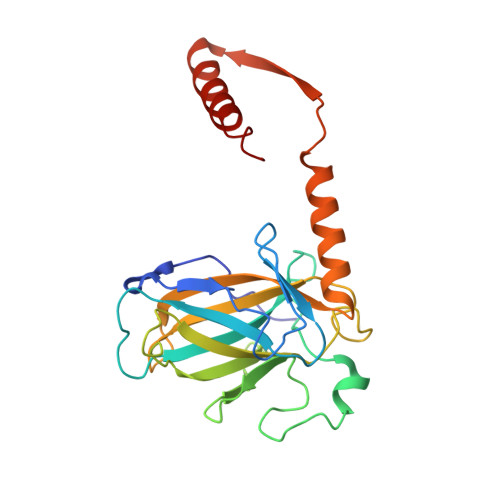
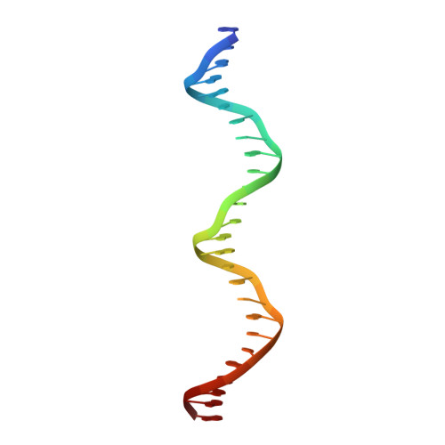
    The p53 tumor suppressor protein is a sequence-specific DNA-binding transcription factor. Structures of p53 bound to DNA have been described, but, so far, no structure has been determined of p53 bound to a natural p53-response element. We describe here the structure of a human p53 homotetramer encompassing both the DNA-binding and homo-oligomerization domains in complex with the natural p53-response element present upstream of the promoter of the CDKN1A (p21) gene. Similar to our previously described structures of human p53 tetramers bound to an artificial consensus DNA site, p53 DNA binding proceeds via an induced fit mechanism with loops L1 of two subunits adopting recessed conformations. Interestingly, the conformational change involving loop L1 is even more extreme than the one previously observed with the artificial consensus DNA site. In fact, the previously determined loop L1 conformation seems to be a transition intermediate between the non-DNA-bound and CDKN1A-bound states. Thus, the new structure further supports our model that recognition of specific DNA by p53 is associated with conformational changes within the DNA-binding domain of p53.


  • Organizational Affiliation

    Department of Molecular Biology, University of Geneva, Geneva, Switzerland.


Macromolecules

Find similar proteins by:  (by identity cutoff)  |  3D Structure
Entity ID: 1
MoleculeChains Sequence LengthOrganismDetailsImage
Cellular tumor antigen p53
A, B, C, D
234Homo sapiensMutation(s): 18 
Gene Names: TP53P53
UniProt & NIH Common Fund Data Resources
Find proteins for P04637 (Homo sapiens)
Explore P04637 
Go to UniProtKB:  P04637
PHAROS:  P04637
GTEx:  ENSG00000141510 
Entity Groups  
Sequence Clusters30% Identity50% Identity70% Identity90% Identity95% Identity100% Identity
UniProt GroupP04637
Sequence Annotations
Expand
  • Reference Sequence
Find similar nucleic acids by:  (by identity cutoff)  |  3D Structure
Entity ID: 2
MoleculeChains LengthOrganismImage
CDKN1A(p21) sense strandE [auth K]26N/A
Sequence Annotations
Expand
  • Reference Sequence
Find similar nucleic acids by:  (by identity cutoff)  |  3D Structure
Entity ID: 3
MoleculeChains LengthOrganismImage
CDKN1A(p21) anti-sense strandF [auth L]26N/A
Sequence Annotations
Expand
  • Reference Sequence
Experimental Data & Validation

Experimental Data

  • Method: X-RAY DIFFRACTION
  • Resolution: 2.80 Å
  • R-Value Free: 0.283 
  • R-Value Work: 0.236 
  • R-Value Observed: 0.236 
  • Space Group: P 21 21 2
Unit Cell:
Length ( Å )Angle ( ˚ )
a = 163.81α = 90
b = 169.203β = 90
c = 55.261γ = 90
Software Package:
Software NamePurpose
MxCuBEdata collection
AMoREphasing
CNSrefinement
MOSFLMdata reduction
SCALAdata scaling

Structure Validation

View Full Validation Report


Entry History 

Deposition Data

Revision History  (Full details and data files)

  • Version 1.0: 2011-11-23
    Type: Initial release
  • Version 1.1: 2017-08-02
    Changes: Refinement description, Source and taxonomy
  • Version 1.2: 2024-02-28
    Changes: Data collection, Database references, Derived calculations