


Crystal structure of the human ubiquitin-conjugating enzyme (E2) UbcH5b
Page, R.C., Amick, J., Misra, S.To be published.
Experimental Data Snapshot
wwPDB Validation 3D Report Full Report

Entity ID: 1 | |||||
|---|---|---|---|---|---|
| Molecule | Chains | Sequence Length | Organism | Details | Image |
| Ubiquitin-conjugating enzyme E2 D2 | 152 | Homo sapiens | Mutation(s): 0 Gene Names: UBC4, UBC5B, UBCH4, UBCH5B, UBE2D2 EC: 6.3.2.19 | 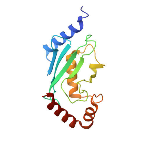 | |
UniProt & NIH Common Fund Data Resources | |||||
Find proteins for P62837 (Homo sapiens) Explore P62837 Go to UniProtKB: P62837 | |||||
PHAROS: P62837 GTEx: ENSG00000131508 | |||||
Entity Groups | |||||
| Sequence Clusters | 30% Identity50% Identity70% Identity90% Identity95% Identity100% Identity | ||||
| UniProt Group | P62837 | ||||
Sequence AnnotationsExpand | |||||
| |||||
| Length ( Å ) | Angle ( ˚ ) |
|---|---|
| a = 49.342 | α = 90 |
| b = 50.277 | β = 90 |
| c = 64.299 | γ = 90 |
| Software Name | Purpose |
|---|---|
| StructureStudio | data collection |
| PHENIX | model building |
| PHENIX | refinement |
| d*TREK | data reduction |
| d*TREK | data scaling |
| PHENIX | phasing |
RCSB PDB (citation) is hosted by
RCSB PDB is a member of the

