3S9N

Complex between transferrin receptor 1 and transferrin with iron in the N-Lobe, room temperature


Experimental Data Snapshot

  • Method: X-RAY DIFFRACTION
  • Resolution: 3.25 Å
  • R-Value Free: 0.289 
  • R-Value Work: 0.254 

wwPDB Validation   3D Report Full Report


Ligand Structure Quality Assessment 


This is version 1.2 of the entry. See complete history


Literature

How the binding of human transferrin primes the transferrin receptor potentiating iron release at endosomal pH.

Eckenroth, B.E.Steere, A.N.Chasteen, N.D.Everse, S.J.Mason, A.B.

(2011) Proc Natl Acad Sci U S A 108: 13089-13094

  • DOI: https://doi.org/10.1073/pnas.1105786108
  • Primary Citation of Related Structures:  
    3S9L, 3S9M, 3S9N

  • PubMed Abstract: 

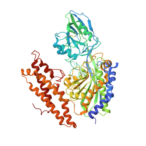
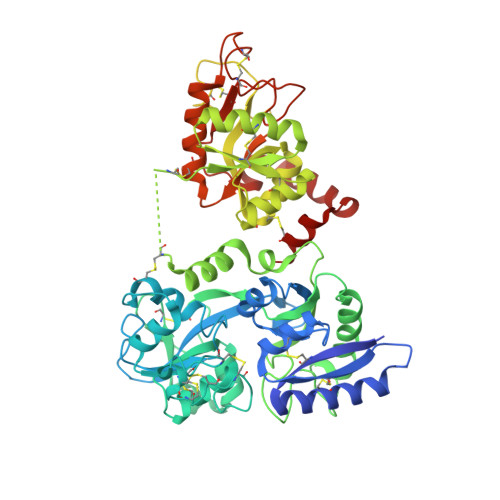
    Delivery of iron to cells requires binding of two iron-containing human transferrin (hTF) molecules to the specific homodimeric transferrin receptor (TFR) on the cell surface. Through receptor-mediated endocytosis involving lower pH, salt, and an unidentified chelator, iron is rapidly released from hTF within the endosome. The crystal structure of a monoferric N-lobe hTF/TFR complex (3.22-Å resolution) features two binding motifs in the N lobe and one in the C lobe of hTF. Binding of Fe(N)hTF induces global and site-specific conformational changes within the TFR ectodomain. Specifically, movements at the TFR dimer interface appear to prime the TFR to undergo pH-induced movements that alter the hTF/TFR interaction. Iron release from each lobe then occurs by distinctly different mechanisms: Binding of His349 to the TFR (strengthened by protonation at low pH) controls iron release from the C lobe, whereas displacement of one N-lobe binding motif, in concert with the action of the dilysine trigger, elicits iron release from the N lobe. One binding motif in each lobe remains attached to the same α-helix in the TFR throughout the endocytic cycle. Collectively, the structure elucidates how the TFR accelerates iron release from the C lobe, slows it from the N lobe, and stabilizes binding of apohTF for return to the cell surface. Importantly, this structure provides new targets for mutagenesis studies to further understand and define this system.


  • Organizational Affiliation

    Department of Biochemistry, University of Vermont, 89 Beaumont Avenue, Burlington, VT 05405, USA.


Macromolecules
Find similar proteins by:  (by identity cutoff)  |  3D Structure
Entity ID: 1
MoleculeChains Sequence LengthOrganismDetailsImage
Transferrin receptor protein 1
A, B
654Homo sapiensMutation(s): 0 
Gene Names: TFR1TFRC
UniProt & NIH Common Fund Data Resources
Find proteins for P02786 (Homo sapiens)
Explore P02786 
Go to UniProtKB:  P02786
PHAROS:  P02786
GTEx:  ENSG00000072274 
Entity Groups  
Sequence Clusters30% Identity50% Identity70% Identity90% Identity95% Identity100% Identity
UniProt GroupP02786
Sequence Annotations
Expand
  • Reference Sequence
Find similar proteins by:  (by identity cutoff)  |  3D Structure
Entity ID: 2
MoleculeChains Sequence LengthOrganismDetailsImage
Serotransferrin
C, D
693Homo sapiensMutation(s): 4 
Gene Names: PRO1400TF
UniProt & NIH Common Fund Data Resources
Find proteins for P02787 (Homo sapiens)
Explore P02787 
Go to UniProtKB:  P02787
PHAROS:  P02787
GTEx:  ENSG00000091513 
Entity Groups  
Sequence Clusters30% Identity50% Identity70% Identity90% Identity95% Identity100% Identity
UniProt GroupP02787
Sequence Annotations
Expand
  • Reference Sequence
Small Molecules
Ligands 4 Unique
IDChains Name / Formula / InChI Key2D Diagram3D Interactions
NAG
Query on NAG

Download Ideal Coordinates CCD File 
E [auth A],
G [auth B]
2-acetamido-2-deoxy-beta-D-glucopyranose
C8 H15 N O6
OVRNDRQMDRJTHS-FMDGEEDCSA-N
CO3
Query on CO3

Download Ideal Coordinates CCD File 
J [auth C],
L [auth D]
CARBONATE ION
C O3
BVKZGUZCCUSVTD-UHFFFAOYSA-L
FE
Query on FE

Download Ideal Coordinates CCD File 
I [auth C],
K [auth D]
FE (III) ION
Fe
VTLYFUHAOXGGBS-UHFFFAOYSA-N
CA
Query on CA

Download Ideal Coordinates CCD File 
F [auth A],
H [auth B]
CALCIUM ION
Ca
BHPQYMZQTOCNFJ-UHFFFAOYSA-N
Experimental Data & Validation

Experimental Data

  • Method: X-RAY DIFFRACTION
  • Resolution: 3.25 Å
  • R-Value Free: 0.289 
  • R-Value Work: 0.254 
  • Space Group: P 43 2 2
Unit Cell:
Length ( Å )Angle ( ˚ )
a = 234.417α = 90
b = 234.417β = 90
c = 169.65γ = 90
Software Package:
Software NamePurpose
DENZOdata reduction
SCALEPACKdata scaling
PHASERphasing
CNSrefinement
PDB_EXTRACTdata extraction

Structure Validation

View Full Validation Report


Ligand Structure Quality Assessment 


Entry History 

Deposition Data

Revision History  (Full details and data files)

  • Version 1.0: 2011-08-10
    Type: Initial release
  • Version 1.1: 2011-12-14
    Changes: Database references
  • Version 1.2: 2020-07-29
    Type: Remediation
    Reason: Carbohydrate remediation
    Changes: Data collection, Database references, Derived calculations, Structure summary