3QB7

Interleukin-4 mutant RGA bound to cytokine receptor common gamma


Experimental Data Snapshot

  • Method: X-RAY DIFFRACTION
  • Resolution: 3.25 Å
  • R-Value Free: 0.292 
  • R-Value Work: 0.231 
  • R-Value Observed: 0.234 

wwPDB Validation   3D Report Full Report


Ligand Structure Quality Assessment 


This is version 2.1 of the entry. See complete history


Literature

Redirecting cell-type specific cytokine responses with engineered interleukin-4 superkines.

Junttila, I.S.Creusot, R.J.Moraga, I.Bates, D.L.Wong, M.T.Alonso, M.N.Suhoski, M.M.Lupardus, P.Meier-Schellersheim, M.Engleman, E.G.Utz, P.J.Fathman, C.G.Paul, W.E.Garcia, K.C.

(2012) Nat Chem Biol 8: 990-998

  • DOI: https://doi.org/10.1038/nchembio.1096
  • Primary Citation of Related Structures:  
    3QB7

  • PubMed Abstract: 

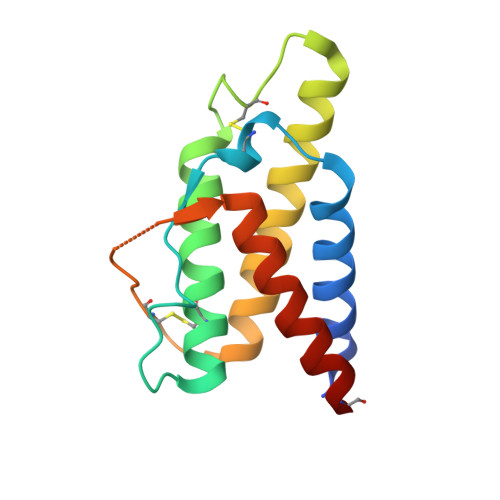
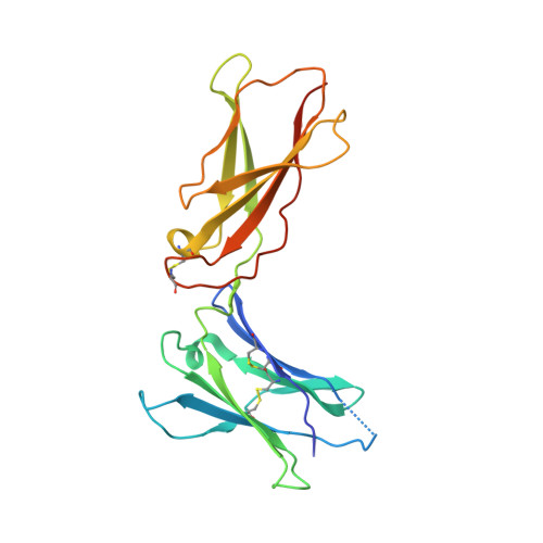
    Cytokines dimerize their receptors, with the binding of the 'second chain' triggering signaling. In the interleukin (IL)-4 and IL-13 system, different cell types express varying numbers of alternative second receptor chains (γc or IL-13Rα1), forming functionally distinct type I or type II complexes. We manipulated the affinity and specificity of second chain recruitment by human IL-4. A type I receptor-selective IL-4 'superkine' with 3,700-fold higher affinity for γc was three- to ten-fold more potent than wild-type IL-4. Conversely, a variant with high affinity for IL-13Rα1 more potently activated cells expressing the type II receptor and induced differentiation of dendritic cells from monocytes, implicating the type II receptor in this process. Superkines showed signaling advantages on cells with lower second chain numbers. Comparative transcriptional analysis reveals that the superkines induce largely redundant gene expression profiles. Variable second chain numbers can be exploited to redirect cytokines toward distinct cell subsets and elicit new actions, potentially improving the selectivity of cytokine therapy.


  • Organizational Affiliation

    Laboratory of Immunology, National Institute of Allergy and Infectious Diseases, National Institutes of Health, Bethesda, Maryland, USA.


Macromolecules
Find similar proteins by:  (by identity cutoff)  |  3D Structure
Entity ID: 1
MoleculeChains Sequence LengthOrganismDetailsImage
Interleukin 4
A, B
132Homo sapiensMutation(s): 8 
Gene Names: IL4
UniProt & NIH Common Fund Data Resources
Find proteins for P05112 (Homo sapiens)
Explore P05112 
Go to UniProtKB:  P05112
PHAROS:  P05112
GTEx:  ENSG00000113520 
Entity Groups  
Sequence Clusters30% Identity50% Identity70% Identity90% Identity95% Identity100% Identity
UniProt GroupP05112
Sequence Annotations
Expand
  • Reference Sequence
Find similar proteins by:  (by identity cutoff)  |  3D Structure
Entity ID: 2
MoleculeChains Sequence LengthOrganismDetailsImage
Cytokine receptor common subunit gamma
C, D
203Homo sapiensMutation(s): 1 
Gene Names: IL2RG
UniProt & NIH Common Fund Data Resources
Find proteins for P31785 (Homo sapiens)
Explore P31785 
Go to UniProtKB:  P31785
PHAROS:  P31785
GTEx:  ENSG00000147168 
Entity Groups  
Sequence Clusters30% Identity50% Identity70% Identity90% Identity95% Identity100% Identity
UniProt GroupP31785
Sequence Annotations
Expand
  • Reference Sequence
Oligosaccharides

Help

Entity ID: 3
MoleculeChains Length2D Diagram Glycosylation3D Interactions
2-acetamido-2-deoxy-beta-D-glucopyranose-(1-4)-2-acetamido-2-deoxy-beta-D-glucopyranose
E, F
2N-Glycosylation
Glycosylation Resources
GlyTouCan:  G42666HT
GlyCosmos:  G42666HT
GlyGen:  G42666HT
Experimental Data & Validation

Experimental Data

  • Method: X-RAY DIFFRACTION
  • Resolution: 3.25 Å
  • R-Value Free: 0.292 
  • R-Value Work: 0.231 
  • R-Value Observed: 0.234 
  • Space Group: P 21 21 21
Unit Cell:
Length ( Å )Angle ( ˚ )
a = 75.969α = 90
b = 105.879β = 90
c = 121.093γ = 90
Software Package:
Software NamePurpose
SCALEPACKdata scaling
PHASERphasing
PHENIXrefinement
PDB_EXTRACTdata extraction
ADSCdata collection
DENZOdata reduction

Structure Validation

View Full Validation Report


Ligand Structure Quality Assessment 


Entry History 

Deposition Data

Revision History  (Full details and data files)

  • Version 1.0: 2012-04-25
    Type: Initial release
  • Version 1.1: 2012-11-14
    Changes: Database references
  • Version 1.2: 2012-12-12
    Changes: Database references
  • Version 1.3: 2017-11-08
    Changes: Advisory, Refinement description
  • Version 2.0: 2020-07-29
    Type: Remediation
    Reason: Carbohydrate remediation
    Changes: Advisory, Atomic model, Data collection, Database references, Derived calculations, Structure summary
  • Version 2.1: 2023-12-27
    Changes: Advisory, Data collection, Database references, Derived calculations, Structure summary