3Q77

Structure of human neutrophil elastase in complex with a dihydropyrimidone inhibitor


Experimental Data Snapshot

  • Method: X-RAY DIFFRACTION
  • Resolution: 2.00 Å
  • R-Value Free: 0.203 
  • R-Value Work: 0.159 
  • R-Value Observed: 0.161 

wwPDB Validation   3D Report Full Report


This is version 2.1 of the entry. See complete history


Literature

Unexpected active-site flexibility in the structure of human neutrophil elastase in complex with a new dihydropyrimidone inhibitor.

Hansen, G.Gielen-Haertwig, H.Reinemer, P.Schomburg, D.Harrenga, A.Niefind, K.

(2011) J Mol Biol 409: 681-691

  • DOI: https://doi.org/10.1016/j.jmb.2011.04.047
  • Primary Citation of Related Structures:  
    3Q76, 3Q77

  • PubMed Abstract: 

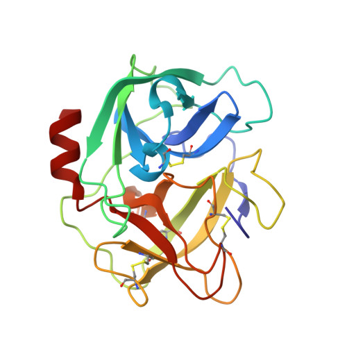
    Human neutrophil elastase (HNE), a trypsin-type serine protease, is of pivotal importance in the onset and progression of chronic obstructive pulmonary disease (COPD). COPD encompasses a group of slowly progressive respiratory disorders and is a major medical problem and the fifth leading cause of death worldwide. HNE is a major target for the development of compounds that inhibit the progression of long-term lung function decline in COPD patients. Here, we present the three-dimensional structure of a potent dihydropyrimidone inhibitor (DHPI) non-covalently bound to HNE at a resolution of 2.0 Å. The inhibitor binds to the active site in a unique orientation addressing S1 and S2 subsites of the protease. To facilitate further analysis of this binding mode, we determined the structure of the uncomplexed enzyme at a resolution of 1.86 Å. Detailed comparisons of the HNE:DHPI complex with the uncomplexed HNE structure and published structures of other elastase:inhibitor complexes revealed that binding of DHPI leads to large conformational changes in residues located in the S2 subsite. The rearrangement of residues Asp95-Leu99B creates a deep, well-defined cavity, which is filled by the P2 moiety of the inhibitor molecule to almost perfect shape complementarity. The shape of the S2 subsite in complex with DHPI clearly differs from all other observed HNE structures. The observed structural flexibility of the S2 subsite is a key feature for the understanding of the binding mode of DHPIs in general and the development of new HNE selective inhibitors.


  • Organizational Affiliation

    Institute of Biochemistry, Department of Chemistry, University of Cologne, Zülpicher Str. 47, D-50674 Cologne, Germany.


Macromolecules
Find similar proteins by:  (by identity cutoff)  |  3D Structure
Entity ID: 1
MoleculeChains Sequence LengthOrganismDetailsImage
Neutrophil elastase218Homo sapiensMutation(s): 0 
EC: 3.4.21.37
UniProt & NIH Common Fund Data Resources
Find proteins for P08246 (Homo sapiens)
Explore P08246 
Go to UniProtKB:  P08246
PHAROS:  P08246
GTEx:  ENSG00000197561 
Entity Groups  
Sequence Clusters30% Identity50% Identity70% Identity90% Identity95% Identity100% Identity
UniProt GroupP08246
Sequence Annotations
Expand
  • Reference Sequence
Oligosaccharides

Help

Entity ID: 2
MoleculeChains Length2D Diagram Glycosylation3D Interactions
2-acetamido-2-deoxy-beta-D-glucopyranose-(1-4)-[alpha-L-fucopyranose-(1-6)]2-acetamido-2-deoxy-beta-D-glucopyranose
B
3N-Glycosylation
Glycosylation Resources
GlyTouCan:  G21290RB
GlyCosmos:  G21290RB
GlyGen:  G21290RB
Small Molecules
Ligands 2 Unique
IDChains Name / Formula / InChI Key2D Diagram3D Interactions
2HY
Query on 2HY

Download Ideal Coordinates CCD File 
D [auth A]2-hydroxyethyl (4R)-4-(4-cyanophenyl)-6-methyl-2-oxo-1-[3-(trifluoromethyl)phenyl]-1,2,3,4-tetrahydropyrimidine-5-carboxylate
C22 H18 F3 N3 O4
XERLAFZWYABQAT-LJQANCHMSA-N
NAG
Query on NAG

Download Ideal Coordinates CCD File 
C [auth A]2-acetamido-2-deoxy-beta-D-glucopyranose
C8 H15 N O6
OVRNDRQMDRJTHS-FMDGEEDCSA-N
Binding Affinity Annotations 
IDSourceBinding Affinity
2HY Binding MOAD:  3Q77 IC50: 6 (nM) from 1 assay(s)
PDBBind:  3Q77 IC50: 6 (nM) from 1 assay(s)
Experimental Data & Validation

Experimental Data

  • Method: X-RAY DIFFRACTION
  • Resolution: 2.00 Å
  • R-Value Free: 0.203 
  • R-Value Work: 0.159 
  • R-Value Observed: 0.161 
  • Space Group: P 63
Unit Cell:
Length ( Å )Angle ( ˚ )
a = 73.732α = 90
b = 73.732β = 90
c = 70.658γ = 120
Software Package:
Software NamePurpose
PHENIXrefinement
AMoREphasing
REFMACrefinement
DENZOdata reduction
SCALEPACKdata scaling
MAR345dtbdata collection

Structure Validation

View Full Validation Report


Entry History 

Deposition Data

Revision History  (Full details and data files)

  • Version 1.0: 2011-05-11
    Type: Initial release
  • Version 1.1: 2011-07-13
    Changes: Version format compliance
  • Version 1.2: 2019-07-17
    Changes: Data collection, Derived calculations, Refinement description
  • Version 2.0: 2020-07-29
    Type: Remediation
    Reason: Carbohydrate remediation
    Changes: Atomic model, Data collection, Derived calculations, Structure summary
  • Version 2.1: 2023-09-13
    Changes: Data collection, Database references, Derived calculations, Refinement description, Structure summary