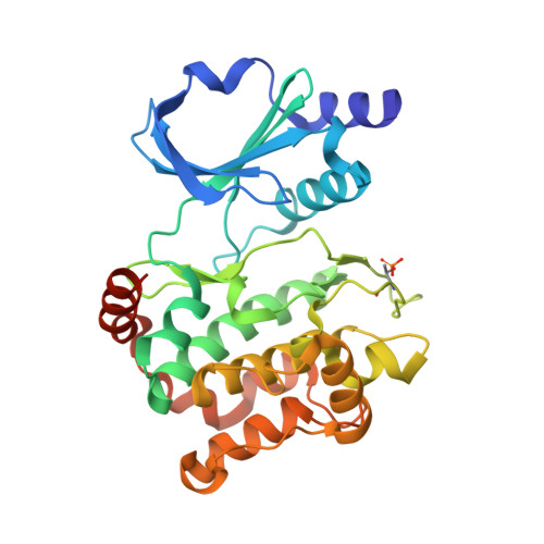
3Q53

Structure of phosphorylated PAK1 kinase domain in complex with ATP


Experimental Data Snapshot

  • Method: X-RAY DIFFRACTION
  • Resolution: 2.09 Å
  • R-Value Free: 0.223 
  • R-Value Work: 0.178 
  • R-Value Observed: 0.180 

wwPDB Validation   3D Report Full Report


This is version 1.2 of the entry. See complete history


Literature

Structural insights into the autoactivation mechanism of p21-activated protein kinase

Wang, J.Wu, J.-W.Wang, Z.-X.

(2011) Structure 19: 1752-1761

  • DOI: https://doi.org/10.1016/j.str.2011.10.013
  • Primary Citation of Related Structures:  
    3Q4Z, 3Q52, 3Q53

  • PubMed Abstract: 

    p21-activated kinases (PAKs) play an important role in diverse cellular processes. Full activation of PAKs requires autophosphorylation of a critical threonine/serine located in the activation loop of the kinase domain. Here we report crystal structures of the phosphorylated and unphosphorylated PAK1 kinase domain. The phosphorylated PAK1 kinase domain has a conformation typical of all active protein kinases. Interestingly, the structure of the unphosphorylated PAK1 kinase domain reveals an unusual dimeric arrangement expected in an authentic enzyme-substrate complex, in which the activation loop of the putative "substrate" is projected into the active site of the "enzyme." The enzyme is bound to AMP-PNP and has an active conformation, whereas the substrate is empty and adopts an inactive conformation. Thus, the structure of the asymmetric homodimer mimics a trans-autophosphorylation complex, and suggests that unphosphorylated PAK1 could dynamically adopt both the active and inactive conformations in solution.


  • Organizational Affiliation

    MOE Key Laboratory for Protein Science, School of Life Sciences, Tsinghua University, Beijing 100084, China.


Macromolecules
Find similar proteins by:  (by identity cutoff)  |  3D Structure
Entity ID: 1
MoleculeChains Sequence LengthOrganismDetailsImage
Serine/threonine-protein kinase PAK 1306Homo sapiensMutation(s): 2 
Gene Names: PAK1
EC: 2.7.11.1
UniProt & NIH Common Fund Data Resources
Find proteins for Q13153 (Homo sapiens)
Explore Q13153 
Go to UniProtKB:  Q13153
PHAROS:  Q13153
GTEx:  ENSG00000149269 
Entity Groups  
Sequence Clusters30% Identity50% Identity70% Identity90% Identity95% Identity100% Identity
UniProt GroupQ13153
Sequence Annotations
Expand
  • Reference Sequence
Small Molecules
Modified Residues  1 Unique
IDChains TypeFormula2D DiagramParent
TPO
Query on TPO
A
L-PEPTIDE LINKINGC4 H10 N O6 PTHR
Experimental Data & Validation

Experimental Data

  • Method: X-RAY DIFFRACTION
  • Resolution: 2.09 Å
  • R-Value Free: 0.223 
  • R-Value Work: 0.178 
  • R-Value Observed: 0.180 
  • Space Group: C 2 2 21
Unit Cell:
Length ( Å )Angle ( ˚ )
a = 48.982α = 90
b = 103.588β = 90
c = 121.967γ = 90
Software Package:
Software NamePurpose
MAR345dtbdata collection
PHASERphasing
PHENIXrefinement
HKL-2000data reduction
HKL-2000data scaling

Structure Validation

View Full Validation Report


Entry History 

Deposition Data

Revision History  (Full details and data files)

  • Version 1.0: 2011-12-21
    Type: Initial release
  • Version 1.1: 2012-04-11
    Changes: Database references
  • Version 1.2: 2023-11-01
    Changes: Data collection, Database references, Derived calculations, Refinement description