3Q01

An induced fit mechanism regulates p53 DNA binding kinetics to confer sequence specificity


Experimental Data Snapshot

  • Method: X-RAY DIFFRACTION
  • Resolution: 2.10 Å
  • R-Value Free: 0.282 
  • R-Value Work: 0.250 
  • R-Value Observed: 0.252 

wwPDB Validation   3D Report Full Report


This is version 1.3 of the entry. See complete history


Literature

An induced fit mechanism regulates p53 DNA binding kinetics to confer sequence specificity.

Petty, T.J.Emamzadah, S.Costantino, L.Petkova, I.Stavridi, E.S.Saven, J.G.Vauthey, E.Halazonetis, T.D.

(2011) EMBO J 30: 2167-2176

  • DOI: https://doi.org/10.1038/emboj.2011.127
  • Primary Citation of Related Structures:  
    3Q01, 3Q05, 3Q06

  • PubMed Abstract: 

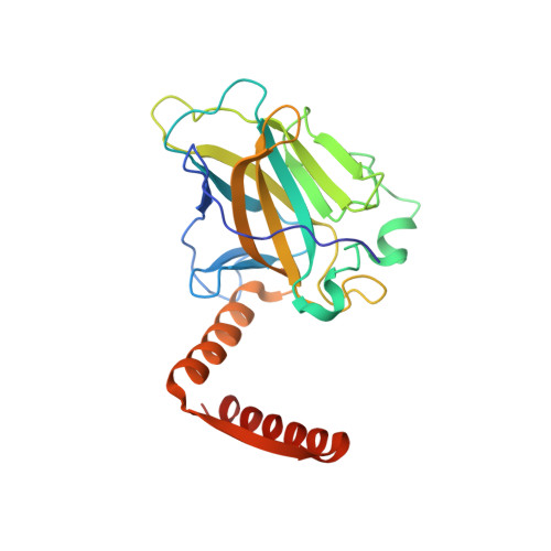
    The p53 tumour suppressor gene, the most frequently mutated gene in human cancer, encodes a transcription factor that contains sequence-specific DNA binding and homo-tetramerization domains. Interestingly, the affinities of p53 for specific and non-specific DNA sites differ by only one order of magnitude, making it hard to understand how this protein recognizes its specific DNA targets in vivo. We describe here the structure of a p53 polypeptide containing both the DNA binding and oligomerization domains in complex with DNA. The structure reveals that sequence-specific DNA binding proceeds via an induced fit mechanism that involves a conformational switch in loop L1 of the p53 DNA binding domain. Analysis of loop L1 mutants demonstrated that the conformational switch allows DNA binding off-rates to be regulated independently of affinities. These results may explain the universal prevalence of conformational switching in sequence-specific DNA binding proteins and suggest that proteins like p53 rely more on differences in binding off-rates, than on differences in affinities, to recognize their specific DNA sites.


  • Organizational Affiliation

    Department of Molecular Biology, University of Geneva, Geneva, Switzerland.


Macromolecules
Find similar proteins by:  (by identity cutoff)  |  3D Structure
Entity ID: 1
MoleculeChains Sequence LengthOrganismDetailsImage
Cellular tumor antigen p53
A, B
233Homo sapiensMutation(s): 17 
Gene Names: P53TP53
UniProt & NIH Common Fund Data Resources
Find proteins for P04637 (Homo sapiens)
Explore P04637 
Go to UniProtKB:  P04637
PHAROS:  P04637
GTEx:  ENSG00000141510 
Entity Groups  
Sequence Clusters30% Identity50% Identity70% Identity90% Identity95% Identity100% Identity
UniProt GroupP04637
Sequence Annotations
Expand
  • Reference Sequence
Experimental Data & Validation

Experimental Data

  • Method: X-RAY DIFFRACTION
  • Resolution: 2.10 Å
  • R-Value Free: 0.282 
  • R-Value Work: 0.250 
  • R-Value Observed: 0.252 
  • Space Group: P 21 21 21
Unit Cell:
Length ( Å )Angle ( ˚ )
a = 69.678α = 90
b = 73.817β = 90
c = 106.685γ = 90
Software Package:
Software NamePurpose
MxCuBEdata collection
AMoREphasing
CNSrefinement
MOSFLMdata reduction
SCALAdata scaling

Structure Validation

View Full Validation Report


Entry History 

Deposition Data

Revision History  (Full details and data files)

  • Version 1.0: 2011-05-11
    Type: Initial release
  • Version 1.1: 2011-07-13
    Changes: Version format compliance
  • Version 1.2: 2017-07-26
    Changes: Refinement description, Source and taxonomy
  • Version 1.3: 2023-09-13
    Changes: Data collection, Database references, Derived calculations, Refinement description