3POM

Crystal Structure of the Unliganded Retinoblastoma Protein Pocket Domain


Experimental Data Snapshot

  • Method: X-RAY DIFFRACTION
  • Resolution: 2.50 Å
  • R-Value Free: 0.262 
  • R-Value Work: 0.213 
  • R-Value Observed: 0.213 

wwPDB Validation   3D Report Full Report


This is version 1.3 of the entry. See complete history


Literature

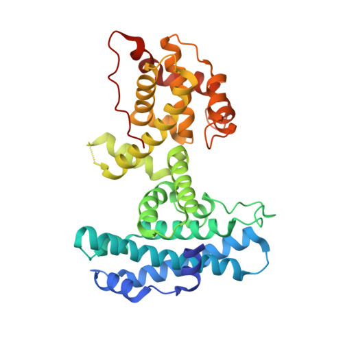
Crystal structure of the unliganded retinoblastoma protein pocket domain.

Balog, E.R.Burke, J.R.Hura, G.L.Rubin, S.M.

(2011) Proteins 79: 2010-2014

  • DOI: https://doi.org/10.1002/prot.23007
  • Primary Citation of Related Structures:  
    3POM

  • PubMed Abstract: 

    The retinoblastoma protein (Rb) regulates cell proliferation through its association with E2F transcription factors and other proteins. The Rb “pocket” domain primarily facilitates protein-protein interactions, and several structures of the pocket bound to E2F and tumorigenic viral proteins have been reported. We report here the first crystal structure of the pocket domain without bound ligand. We find that ligand association results in observable structural changes at the binding sites but no significant changes to the overall conformation of the domain. These data support models for regulation of Rb-E2F binding that do not require considerable structural changes in the pocket domain.


  • Organizational Affiliation

    Department of Molecular, Cell, and Developmental Biology, University of California, Santa Cruz, CA 95064, USA.


Macromolecules
Find similar proteins by:  (by identity cutoff)  |  3D Structure
Entity ID: 1
MoleculeChains Sequence LengthOrganismDetailsImage
Retinoblastoma-associated protein
A, B
352Homo sapiensMutation(s): 0 
Gene Names: RB1
UniProt & NIH Common Fund Data Resources
Find proteins for P06400 (Homo sapiens)
Explore P06400 
Go to UniProtKB:  P06400
PHAROS:  P06400
GTEx:  ENSG00000139687 
Entity Groups  
Sequence Clusters30% Identity50% Identity70% Identity90% Identity95% Identity100% Identity
UniProt GroupP06400
Sequence Annotations
Expand
  • Reference Sequence
Experimental Data & Validation

Experimental Data

  • Method: X-RAY DIFFRACTION
  • Resolution: 2.50 Å
  • R-Value Free: 0.262 
  • R-Value Work: 0.213 
  • R-Value Observed: 0.213 
  • Space Group: P 21 21 21
Unit Cell:
Length ( Å )Angle ( ˚ )
a = 60.282α = 90
b = 111.164β = 90
c = 138.93γ = 90
Software Package:
Software NamePurpose
Blu-Icedata collection
PHASERphasing
PHENIXrefinement
MOSFLMdata reduction
SCALAdata scaling

Structure Validation

View Full Validation Report


Entry History 

Deposition Data

Revision History  (Full details and data files)

  • Version 1.0: 2011-04-27
    Type: Initial release
  • Version 1.1: 2011-07-13
    Changes: Version format compliance
  • Version 1.2: 2017-08-09
    Changes: Refinement description, Source and taxonomy
  • Version 1.3: 2017-11-08
    Changes: Refinement description