3PE4

Structure of human O-GlcNAc transferase and its complex with a peptide substrate


Experimental Data Snapshot

  • Method: X-RAY DIFFRACTION
  • Resolution: 1.95 Å
  • R-Value Free: 0.252 
  • R-Value Work: 0.224 
  • R-Value Observed: 0.225 

wwPDB Validation   3D Report Full Report


Ligand Structure Quality Assessment 


This is version 1.2 of the entry. See complete history


Literature

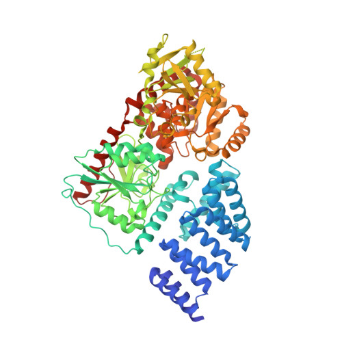
Structure of human O-GlcNAc transferase and its complex with a peptide substrate.

Lazarus, M.B.Nam, Y.Jiang, J.Sliz, P.Walker, S.

(2011) Nature 469: 564-567

  • DOI: https://doi.org/10.1038/nature09638
  • Primary Citation of Related Structures:  
    3PE3, 3PE4

  • PubMed Abstract: 

    The essential mammalian enzyme O-linked β-N-acetylglucosamine transferase (O-GlcNAc transferase, here OGT) couples metabolic status to the regulation of a wide variety of cellular signalling pathways by acting as a nutrient sensor. OGT catalyses the transfer of N-acetylglucosamine from UDP-N-acetylglucosamine (UDP-GlcNAc) to serines and threonines of cytoplasmic, nuclear and mitochondrial proteins, including numerous transcription factors, tumour suppressors, kinases, phosphatases and histone-modifying proteins. Aberrant glycosylation by OGT has been linked to insulin resistance, diabetic complications, cancer and neurodegenerative diseases including Alzheimer's. Despite the importance of OGT, the details of how it recognizes and glycosylates its protein substrates are largely unknown. We report here two crystal structures of human OGT, as a binary complex with UDP (2.8 Å resolution) and as a ternary complex with UDP and a peptide substrate (1.95 Å). The structures provide clues to the enzyme mechanism, show how OGT recognizes target peptide sequences, and reveal the fold of the unique domain between the two halves of the catalytic region. This information will accelerate the rational design of biological experiments to investigate OGT's functions; it will also help the design of inhibitors for use as cellular probes and help to assess its potential as a therapeutic target.


  • Organizational Affiliation

    Department of Chemistry and Chemical Biology, Harvard University, Cambridge, Massachusetts 02138, USA.


Macromolecules
Find similar proteins by:  (by identity cutoff)  |  3D Structure
Entity ID: 1
MoleculeChains Sequence LengthOrganismDetailsImage
UDP-N-acetylglucosamine--peptide N-acetylglucosaminyltransferase 110 kDa subunit
A, C
723Homo sapiensMutation(s): 0 
Gene Names: OGT
EC: 2.4.1
UniProt & NIH Common Fund Data Resources
Find proteins for O15294 (Homo sapiens)
Explore O15294 
Go to UniProtKB:  O15294
PHAROS:  O15294
GTEx:  ENSG00000147162 
Entity Groups  
Sequence Clusters30% Identity50% Identity70% Identity90% Identity95% Identity100% Identity
UniProt GroupO15294
Sequence Annotations
Expand
  • Reference Sequence

Find similar proteins by:  Sequence   |   3D Structure  

Entity ID: 2
MoleculeChains Sequence LengthOrganismDetailsImage
Casein kinase II subunit alpha
B, D
14Homo sapiensMutation(s): 0 
UniProt & NIH Common Fund Data Resources
Find proteins for P68400 (Homo sapiens)
Explore P68400 
Go to UniProtKB:  P68400
PHAROS:  P68400
GTEx:  ENSG00000101266 
Entity Groups  
Sequence Clusters30% Identity50% Identity70% Identity90% Identity95% Identity100% Identity
UniProt GroupP68400
Sequence Annotations
Expand
  • Reference Sequence
Experimental Data & Validation

Experimental Data

Unit Cell:
Length ( Å )Angle ( ˚ )
a = 98.6α = 90
b = 136.7β = 102.9
c = 153.5γ = 90
Software Package:
Software NamePurpose
ADSCdata collection
PHASERphasing
PHENIXrefinement
iMOSFLMdata reduction
SCALAdata scaling

Structure Validation

View Full Validation Report


Ligand Structure Quality Assessment 


Entry History 

Deposition Data

Revision History  (Full details and data files)

  • Version 1.0: 2011-01-19
    Type: Initial release
  • Version 1.1: 2011-07-13
    Changes: Version format compliance
  • Version 1.2: 2024-02-21
    Changes: Data collection, Database references, Derived calculations