3OTB

Crystal structure of human tRNAHis guanylyltransferase (Thg1) - dGTP complex


Experimental Data Snapshot

  • Method: X-RAY DIFFRACTION
  • Resolution: 2.95 Å
  • R-Value Free: 0.301 
  • R-Value Work: 0.254 

wwPDB Validation   3D Report Full Report


This is version 1.3 of the entry. See complete history


Literature

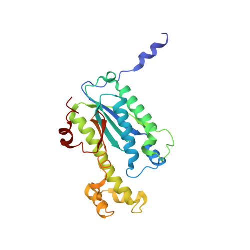
tRNAHis guanylyltransferase (THG1), a unique 3'-5' nucleotidyl transferase, shares unexpected structural homology with canonical 5'-3' DNA polymerases.

Hyde, S.J.Eckenroth, B.E.Smith, B.A.Eberley, W.A.Heintz, N.H.Jackman, J.E.Doublie, S.

(2010) Proc Natl Acad Sci U S A 107: 20305-20310

  • DOI: https://doi.org/10.1073/pnas.1010436107
  • Primary Citation of Related Structures:  
    3OTB, 3OTC, 3OTD, 3OTE

  • PubMed Abstract: 

    All known DNA and RNA polymerases catalyze the formation of phosphodiester bonds in a 5' to 3' direction, suggesting this property is a fundamental feature of maintaining and dispersing genetic information. The tRNA(His) guanylyltransferase (Thg1) is a member of a unique enzyme family whose members catalyze an unprecedented reaction in biology: 3'-5' addition of nucleotides to nucleic acid substrates. The 2.3-Å crystal structure of human THG1 (hTHG1) reported here shows that, despite the lack of sequence similarity, hTHG1 shares unexpected structural homology with canonical 5'-3' DNA polymerases and adenylyl/guanylyl cyclases, two enzyme families known to use a two-metal-ion mechanism for catalysis. The ability of the same structural architecture to catalyze both 5'-3' and 3'-5' reactions raises important questions concerning selection of the 5'-3' mechanism during the evolution of nucleotide polymerases.


  • Organizational Affiliation

    Department of Microbiology and Molecular Genetics, University of Vermont, Burlington, Vermont 05405, USA.


Macromolecules
Find similar proteins by:  (by identity cutoff)  |  3D Structure
Entity ID: 1
MoleculeChains Sequence LengthOrganismDetailsImage
tRNA(His) guanylyltransferase
A, B
269Homo sapiensMutation(s): 0 
Gene Names: ICF45THG1L
EC: 2.7.7
UniProt & NIH Common Fund Data Resources
Find proteins for Q9NWX6 (Homo sapiens)
Explore Q9NWX6 
Go to UniProtKB:  Q9NWX6
PHAROS:  Q9NWX6
GTEx:  ENSG00000113272 
Entity Groups  
Sequence Clusters30% Identity50% Identity70% Identity90% Identity95% Identity100% Identity
UniProt GroupQ9NWX6
Sequence Annotations
Expand
  • Reference Sequence
Small Molecules
Ligands 3 Unique
IDChains Name / Formula / InChI Key2D Diagram3D Interactions
DGT
Query on DGT

Download Ideal Coordinates CCD File 
C [auth A],
I [auth B]
2'-DEOXYGUANOSINE-5'-TRIPHOSPHATE
C10 H16 N5 O13 P3
HAAZLUGHYHWQIW-KVQBGUIXSA-N
3PO
Query on 3PO

Download Ideal Coordinates CCD File 
G [auth A],
H [auth B]
TRIPHOSPHATE
H5 O10 P3
UNXRWKVEANCORM-UHFFFAOYSA-N
MG
Query on MG

Download Ideal Coordinates CCD File 
D [auth A]
E [auth A]
F [auth A]
J [auth B]
K [auth B]
D [auth A],
E [auth A],
F [auth A],
J [auth B],
K [auth B],
L [auth B]
MAGNESIUM ION
Mg
JLVVSXFLKOJNIY-UHFFFAOYSA-N
Experimental Data & Validation

Experimental Data

  • Method: X-RAY DIFFRACTION
  • Resolution: 2.95 Å
  • R-Value Free: 0.301 
  • R-Value Work: 0.254 
  • Space Group: P 43 21 2
Unit Cell:
Length ( Å )Angle ( ˚ )
a = 140.912α = 90
b = 140.912β = 90
c = 81.227γ = 90
Software Package:
Software NamePurpose
DENZOdata reduction
SCALEPACKdata scaling
MOLREPphasing
CNSrefinement
PDB_EXTRACTdata extraction

Structure Validation

View Full Validation Report


Entry History 

Deposition Data

Revision History  (Full details and data files)

  • Version 1.0: 2010-11-17
    Type: Initial release
  • Version 1.1: 2011-07-13
    Changes: Version format compliance
  • Version 1.2: 2014-04-09
    Changes: Source and taxonomy
  • Version 1.3: 2024-02-21
    Changes: Data collection, Database references, Derived calculations