3OQS

Crystal structure of importin-alpha bound to a CLIC4 NLS peptide


Experimental Data Snapshot

  • Method: X-RAY DIFFRACTION
  • Resolution: 2.00 Å
  • R-Value Free: 0.237 
  • R-Value Work: 0.197 
  • R-Value Observed: 0.199 

wwPDB Validation   3D Report Full Report


This is version 1.1 of the entry. See complete history


Literature

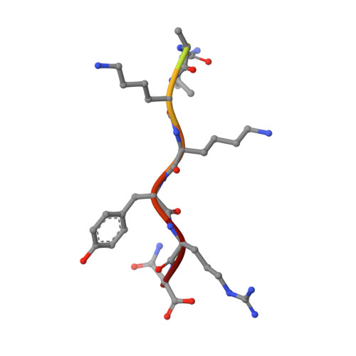
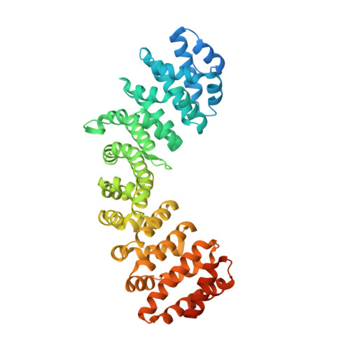
Crystal structure of importin-alpha bound to a peptide bearing the nuclear localisation signal from chloride intracellular channel protein 4

Mynott, A.V.Harrop, S.J.Brown, L.J.Breit, S.N.Kobe, B.Curmi, P.M.G.

(2011) FEBS J 278: 1662-1675

  • DOI: https://doi.org/10.1111/j.1742-4658.2011.08086.x
  • Primary Citation of Related Structures:  
    3OQS

  • PubMed Abstract: 

    It has been reported that a human chloride intracellular channel (CLIC) protein, CLIC4, translocates to the nucleus in response to cellular stress, facilitated by a putative CLIC4 nuclear localization signal (NLS). The CLIC4 NLS adopts an α-helical structure in the native CLIC4 fold. It is proposed that CLIC4 is transported to the nucleus via the classical nuclear import pathway after binding the import receptor, importin-α. In this study, we have determined the X-ray crystal structure of a truncated form of importin-α lacking the importin-β binding domain, bound to a CLIC4 NLS peptide. The NLS peptide binds to the major binding site in an extended conformation similar to that observed for the classical simian virus 40 large T-antigen NLS. A Tyr residue within the CLIC4 NLS makes surprisingly favourable interactions by forming side-chain hydrogen bonds to the importin-α backbone. This structural evidence supports the hypothesis that CLIC4 translocation to the nucleus is governed by the importin-α nuclear import pathway, provided that CLIC4 can undergo a conformational rearrangement that exposes the NLS in an extended conformation.


  • Organizational Affiliation

    School of Physics, University of New South Wales, Sydney, NSW, Australia.


Macromolecules

Find similar proteins by:  Sequence   |   3D Structure  

Entity ID: 1
MoleculeChains Sequence LengthOrganismDetailsImage
peptide of Chloride intracellular channel protein 4A [auth B]10Homo sapiensMutation(s): 0 
UniProt & NIH Common Fund Data Resources
Find proteins for Q9Y696 (Homo sapiens)
Explore Q9Y696 
Go to UniProtKB:  Q9Y696
PHAROS:  Q9Y696
GTEx:  ENSG00000169504 
Entity Groups  
Sequence Clusters30% Identity50% Identity70% Identity90% Identity95% Identity100% Identity
UniProt GroupQ9Y696
Sequence Annotations
Expand
  • Reference Sequence
Find similar proteins by:  (by identity cutoff)  |  3D Structure
Entity ID: 2
MoleculeChains Sequence LengthOrganismDetailsImage
Importin subunit alpha-2B [auth A]510Mus musculusMutation(s): 0 
Gene Names: Kpna2Rch1
UniProt & NIH Common Fund Data Resources
Find proteins for P52293 (Mus musculus)
Explore P52293 
Go to UniProtKB:  P52293
IMPC:  MGI:103561
Entity Groups  
Sequence Clusters30% Identity50% Identity70% Identity90% Identity95% Identity100% Identity
UniProt GroupP52293
Sequence Annotations
Expand
  • Reference Sequence
Experimental Data & Validation

Experimental Data

  • Method: X-RAY DIFFRACTION
  • Resolution: 2.00 Å
  • R-Value Free: 0.237 
  • R-Value Work: 0.197 
  • R-Value Observed: 0.199 
  • Space Group: P 21 21 21
Unit Cell:
Length ( Å )Angle ( ˚ )
a = 78.594α = 90
b = 89.558β = 90
c = 100.083γ = 90
Software Package:
Software NamePurpose
MOSFLMdata reduction
SCALAdata scaling
REFMACrefinement
PDB_EXTRACTdata extraction

Structure Validation

View Full Validation Report


Entry History 

Deposition Data

Revision History  (Full details and data files)

  • Version 1.0: 2011-07-27
    Type: Initial release
  • Version 1.1: 2023-11-01
    Changes: Data collection, Database references, Refinement description