3O78

The structure of Ca2+ Sensor (Case-12)


Experimental Data Snapshot

  • Method: X-RAY DIFFRACTION
  • Resolution: 2.60 Å
  • R-Value Free: 0.316 
  • R-Value Work: 0.242 
  • R-Value Observed: 0.245 

wwPDB Validation   3D Report Full Report


This is version 1.6 of the entry. See complete history


Literature

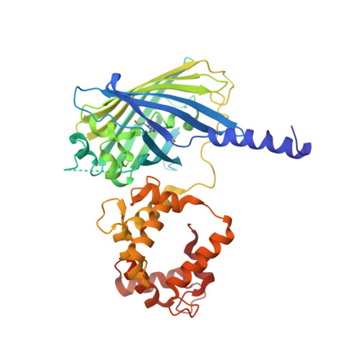
The structure of Ca2+ sensor Case16 reveals the mechanism of reaction to low Ca2+ concentrations

Leder, L.Stark, W.Freuler, F.Marsh, M.Meyerhofer, M.Stettler, T.Mayr, L.M.Britanova, O.V.Strukova, L.A.Chudakov, D.M.Souslova, E.A.

(2010) Sensors (Basel) 10: 8143-8160

  • DOI: https://doi.org/10.3390/s100908143
  • Primary Citation of Related Structures:  
    3O77, 3O78

  • PubMed Abstract: 

    Here we report the first crystal structure of a high-contrast genetically encoded circularly permuted green fluorescent protein (cpGFP)-based Ca(2+) sensor, Case16, in the presence of a low Ca(2+) concentration. The structure reveals the positioning of the chromophore within Case16 at the first stage of the Ca(2+)-dependent response when only two out of four Ca(2+)-binding pockets of calmodulin (CaM) are occupied with Ca(2+) ions. In such a "half Ca(2+)-bound state", Case16 is characterized by an incomplete interaction between its CaM-/M13-domains. We also report the crystal structure of the related Ca(2+) sensor Case12 at saturating Ca(2+) concentration. Based on this structure, we postulate that cpGFP-based Ca(2+) sensors can form non-functional homodimers where the CaM-domain of one sensor molecule binds symmetrically to the M13-peptide of the partner sensor molecule. Case12 and Case16 behavior upon addition of high concentrations of free CaM or M13-peptide reveals that the latter effectively blocks the fluorescent response of the sensor. We speculate that the demonstrated intermolecular interaction with endogenous substrates and homodimerization can impede proper functioning of this type of Ca(2+) sensors in living cells.


  • Organizational Affiliation

    Novartis Pharma AG, NIBR/CPC/LFP, Lichtstrasse 35, CH-4002 Basel, Switzerland. lukas.leder@novartis.com


Macromolecules
Find similar proteins by:  (by identity cutoff)  |  3D Structure
Entity ID: 1
MoleculeChains Sequence LengthOrganismDetailsImage
Myosin light chain kinase, smooth muscle,Green fluorescent protein,Green fluorescent protein,Calmodulin-1
A, B
415Gallus gallusAequorea victoriaHomo sapiensMutation(s): 18 
Gene Names: MylkGFPCALM1CALMCAMCAM1
EC: 2.7.11.18
UniProt & NIH Common Fund Data Resources
Find proteins for P42212 (Aequorea victoria)
Explore P42212 
Go to UniProtKB:  P42212
Find proteins for P0DP23 (Homo sapiens)
Explore P0DP23 
Go to UniProtKB:  P0DP23
PHAROS:  P0DP23
GTEx:  ENSG00000198668 
Find proteins for P11799 (Gallus gallus)
Explore P11799 
Go to UniProtKB:  P11799
Entity Groups  
Sequence Clusters30% Identity50% Identity70% Identity90% Identity95% Identity100% Identity
UniProt GroupsP0DP23P42212P11799
Sequence Annotations
Expand
  • Reference Sequence
Experimental Data & Validation

Experimental Data

  • Method: X-RAY DIFFRACTION
  • Resolution: 2.60 Å
  • R-Value Free: 0.316 
  • R-Value Work: 0.242 
  • R-Value Observed: 0.245 
  • Space Group: P 1 21 1
Unit Cell:
Length ( Å )Angle ( ˚ )
a = 46.374α = 90
b = 101.635β = 91.45
c = 82.211γ = 90
Software Package:
Software NamePurpose
MAR345dtbdata collection
MOLREPphasing
REFMACrefinement
XDSdata reduction
XSCALEdata scaling

Structure Validation

View Full Validation Report


Entry History 

Deposition Data

Revision History  (Full details and data files)

  • Version 1.0: 2010-09-29
    Type: Initial release
  • Version 1.1: 2011-07-13
    Changes: Version format compliance
  • Version 1.2: 2012-04-25
    Changes: Database references
  • Version 1.3: 2012-06-13
    Changes: Database references
  • Version 1.4: 2017-06-21
    Changes: Data collection, Database references, Source and taxonomy, Structure summary
  • Version 1.5: 2023-11-01
    Changes: Advisory, Data collection, Database references, Derived calculations, Refinement description
  • Version 1.6: 2023-12-06
    Changes: Data collection