3O3Q

Crystal structure of "L44F/M67I/L73V/A103G/deletion 104-106/F108Y/V109L/L111I/C117V/R119G/deletion 120-122" mutant form of Human acidic fibroblast growth factor


Experimental Data Snapshot

  • Method: X-RAY DIFFRACTION
  • Resolution: 1.60 Å
  • R-Value Free: 0.203 
  • R-Value Work: 0.170 
  • R-Value Observed: 0.172 

wwPDB Validation   3D Report Full Report


This is version 1.4 of the entry. See complete history


Literature

A polypeptide "building block"top-down symmetric deconstruction".

Lee, J.Blaber, S.I.Dubey, V.K.Blaber, M.

(2011) J Mol Biol 407: 744-763

  • DOI: https://doi.org/10.1016/j.jmb.2011.02.002
  • Primary Citation of Related Structures:  
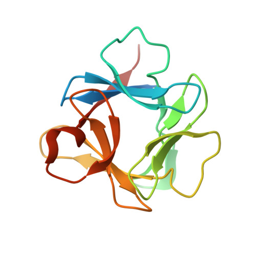
    3O3Q

  • PubMed Abstract: 

    Fibroblast growth factor-1, a member of the 3-fold symmetric β-trefoil fold, was subjected to a series of symmetric constraint mutations in a process termed "top-down symmetric deconstruction." The mutations enforced a cumulative exact 3-fold symmetry upon symmetrically equivalent positions within the protein and were combined with a stability screen. This process culminated in a β-trefoil protein with exact 3-fold primary-structure symmetry that exhibited excellent folding and stability properties. Subsequent fragmentation of the repeating primary-structure motif yielded a 42-residue polypeptide capable of spontaneous assembly as a homotrimer, producing a thermostable β-trefoil architecture. The results show that despite pronounced reduction in sequence complexity, pure symmetry in the design of a foldable, thermostable β-trefoil fold is possible. The top-down symmetric deconstruction approach provides a novel alternative means to successfully identify a useful polypeptide "building block" for subsequent "bottom-up" de novo design of target protein architecture.


  • Organizational Affiliation

    Department of Biomedical Sciences, College of Medicine, Florida State University, Tallahassee, FL 32306-4300, USA.


Macromolecules
Find similar proteins by:  (by identity cutoff)  |  3D Structure
Entity ID: 1
MoleculeChains Sequence LengthOrganismDetailsImage
Heparin-binding growth factor 1
A, B, C, D
140Homo sapiensMutation(s): 9 
Gene Names: FGF1FGFA
UniProt & NIH Common Fund Data Resources
Find proteins for P05230 (Homo sapiens)
Explore P05230 
Go to UniProtKB:  P05230
PHAROS:  P05230
GTEx:  ENSG00000113578 
Entity Groups  
Sequence Clusters30% Identity50% Identity70% Identity90% Identity95% Identity100% Identity
UniProt GroupP05230
Sequence Annotations
Expand
  • Reference Sequence
Experimental Data & Validation

Experimental Data

  • Method: X-RAY DIFFRACTION
  • Resolution: 1.60 Å
  • R-Value Free: 0.203 
  • R-Value Work: 0.170 
  • R-Value Observed: 0.172 
  • Space Group: P 1
Unit Cell:
Length ( Å )Angle ( ˚ )
a = 46.92α = 64.94
b = 56.89β = 89.76
c = 61.92γ = 71.27
Software Package:
Software NamePurpose
PHENIXrefinement
CNSrefinement
HKL-2000data collection
HKL-2000data reduction
HKL-2000data scaling
CNSphasing

Structure Validation

View Full Validation Report


Entry History 

Deposition Data

Revision History  (Full details and data files)

  • Version 1.0: 2011-02-23
    Type: Initial release
  • Version 1.1: 2011-07-13
    Changes: Version format compliance
  • Version 1.2: 2012-06-20
    Changes: Database references
  • Version 1.3: 2019-07-17
    Changes: Data collection, Refinement description
  • Version 1.4: 2023-09-06
    Changes: Data collection, Database references, Derived calculations, Refinement description