


Structure-function analysis of human L-Prostaglandin D Synthase bound with fatty acid
Zhou, Y., Shaw, N., Li, Y., Zhao, Y., Zhang, R., Liu, Z.-J.To be published.
Experimental Data Snapshot
Entity ID: 1 | |||||
|---|---|---|---|---|---|
| Molecule | Chains | Sequence Length | Organism | Details | Image |
| Prostaglandin-H2 D-isomerase | 162 | Homo sapiens | Mutation(s): 1 EC: 5.3.99.2 | 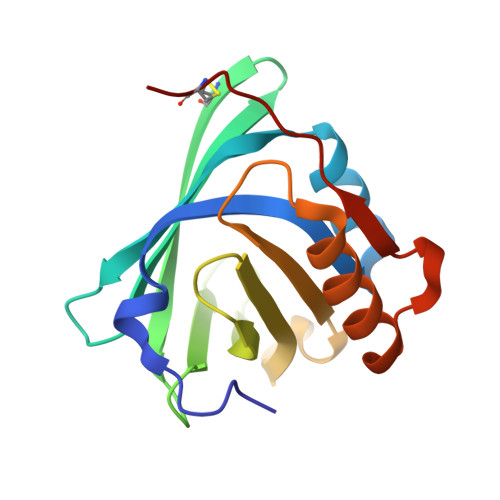 | |
UniProt & NIH Common Fund Data Resources | |||||
Find proteins for P41222 (Homo sapiens) Explore P41222 Go to UniProtKB: P41222 | |||||
PHAROS: P41222 GTEx: ENSG00000107317 | |||||
Entity Groups | |||||
| Sequence Clusters | 30% Identity50% Identity70% Identity90% Identity95% Identity100% Identity | ||||
| UniProt Group | P41222 | ||||
Sequence AnnotationsExpand | |||||
| |||||
| Ligands 3 Unique | |||||
|---|---|---|---|---|---|
| ID | Chains | Name / Formula / InChI Key | 2D Diagram | 3D Interactions | |
| OLA Query on OLA | C [auth A], E [auth B] | OLEIC ACID C18 H34 O2 ZQPPMHVWECSIRJ-KTKRTIGZSA-N |  | ||
| PLM Query on PLM | D [auth A], F [auth B] | PALMITIC ACID C16 H32 O2 IPCSVZSSVZVIGE-UHFFFAOYSA-N |  | ||
| GOL Query on GOL | G [auth B] | GLYCEROL C3 H8 O3 PEDCQBHIVMGVHV-UHFFFAOYSA-N |  | ||
| Length ( Å ) | Angle ( ˚ ) |
|---|---|
| a = 90.221 | α = 90 |
| b = 90.221 | β = 90 |
| c = 35.635 | γ = 90 |
| Software Name | Purpose |
|---|---|
| HKL-2000 | data collection |
| PHASES | phasing |
| REFMAC | refinement |
| HKL-2000 | data reduction |
| HKL-2000 | data scaling |


