3N3F

Crystal Structure of the Human Collagen XV Trimerization Domain: A Potent Trimerizing Unit Common to Multiplexin Collagens


Experimental Data Snapshot

  • Method: X-RAY DIFFRACTION
  • Resolution: 2.00 Å
  • R-Value Free: 0.250 
  • R-Value Work: 0.216 

wwPDB Validation   3D Report Full Report


This is version 1.2 of the entry. See complete history


Literature

Crystal structure of the human collagen XV trimerization domain: A potent trimerizing unit common to multiplexin collagens.

Wirz, J.A.Boudko, S.P.Lerch, T.F.Chapman, M.S.Bachinger, H.P.

(2011) Matrix Biol 30: 9-15

  • DOI: https://doi.org/10.1016/j.matbio.2010.09.005
  • Primary Citation of Related Structures:  
    3N3F

  • PubMed Abstract: 

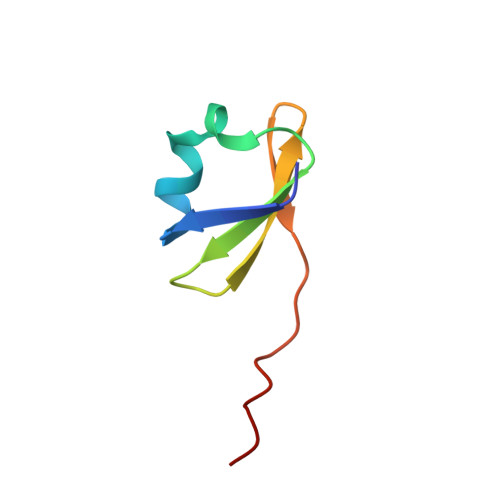
    Correct folding of the collagen triple helix requires a self-association step which selects and binds α-chains into trimers. Here we report the crystal structure of the trimerization domain of human type XV collagen. The trimerization domain of type XV collagen contains three monomers each composed of four β-sheets and an α-helix. The hydrophobic core of the trimer is devoid of solvent molecules and is shaped by β-sheet planes from each monomer. The trimerization domain is extremely stable and forms at picomolar concentrations. It is found that the trimerization domain of type XV collagen is structurally similar to that of type XVIII, despite only 32% sequence identity. High structural conservation indicates that the multiplexin trimerization domain represents a three dimensional fold that allows for sequence variability while retaining structural integrity necessary for tight and efficient trimerization.


  • Organizational Affiliation

    Research Department of Shriners Hospital for Children, Portland, OR 97239, USA.


Macromolecules
Find similar proteins by:  (by identity cutoff)  |  3D Structure
Entity ID: 1
MoleculeChains Sequence LengthOrganismDetailsImage
Collagen alpha-1(XV) chain
A, B
54Homo sapiensMutation(s): 0 
Gene Names: COL15A1
UniProt & NIH Common Fund Data Resources
Find proteins for P39059 (Homo sapiens)
Explore P39059 
Go to UniProtKB:  P39059
PHAROS:  P39059
GTEx:  ENSG00000204291 
Entity Groups  
Sequence Clusters30% Identity50% Identity70% Identity90% Identity95% Identity100% Identity
UniProt GroupP39059
Sequence Annotations
Expand
  • Reference Sequence
Experimental Data & Validation

Experimental Data

  • Method: X-RAY DIFFRACTION
  • Resolution: 2.00 Å
  • R-Value Free: 0.250 
  • R-Value Work: 0.216 
  • Space Group: P 63
Unit Cell:
Length ( Å )Angle ( ˚ )
a = 49.12α = 90
b = 49.12β = 90
c = 65.6γ = 120
Software Package:
Software NamePurpose
MOSFLMdata reduction
SCALAdata scaling
AMoREphasing
CNSrefinement
PDB_EXTRACTdata extraction
CrystalCleardata collection

Structure Validation

View Full Validation Report


Entry History 

Deposition Data

  • Released Date: 2010-10-20 
  • Deposition Author(s): Wirz, J.A.

Revision History  (Full details and data files)

  • Version 1.0: 2010-10-20
    Type: Initial release
  • Version 1.1: 2011-07-13
    Changes: Version format compliance
  • Version 1.2: 2023-09-06
    Changes: Data collection, Database references, Refinement description