3N2S

Structure of NfrA1 nitroreductase from B. subtilis


Experimental Data Snapshot

  • Method: X-RAY DIFFRACTION
  • Resolution: 1.95 Å
  • R-Value Free: 0.198 
  • R-Value Work: 0.173 
  • R-Value Observed: 0.174 

wwPDB Validation   3D Report Full Report


Ligand Structure Quality Assessment 


This is version 1.1 of the entry. See complete history


Literature

NADH oxidase activity of Bacillus subtilis nitroreductase NfrA1: insight into its biological role.

Cortial, S.Chaignon, P.Iorga, B.I.Aymerich, S.Truan, G.Gueguen-Chaignon, V.Meyer, P.Morera, S.Ouazzani, J.

(2010) FEBS Lett 584: 3916-3922

  • DOI: https://doi.org/10.1016/j.febslet.2010.08.019
  • Primary Citation of Related Structures:  
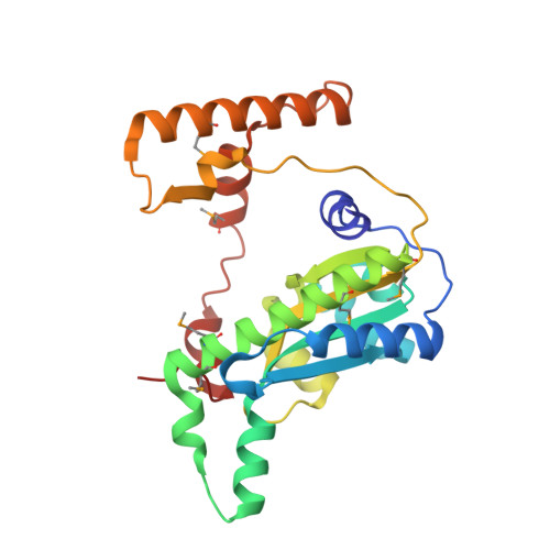
    3N2S

  • PubMed Abstract: 

    NfrA1 nitroreductase from the Gram-positive bacterium Bacillus subtilis is a member of the NAD(P)H/FMN oxidoreductase family. Here, we investigated the reactivity, the structure and kinetics of NfrA1, which could provide insight into the unclear biological role of this enzyme. We could show that NfrA1 possesses an NADH oxidase activity that leads to high concentrations of oxygen peroxide and an NAD(+) degrading activity leading to free nicotinamide. Finally, we showed that NfrA1 is able to rapidly scavenge H(2)O(2) produced during the oxidative process or added exogenously.


  • Organizational Affiliation

    Institut de Chimie des Substances Naturelles (ICSN), CNRS UPR 2301, Gif-sur-Yvette, France.


Macromolecules
Find similar proteins by:  (by identity cutoff)  |  3D Structure
Entity ID: 1
MoleculeChains Sequence LengthOrganismDetailsImage
NADPH-dependent nitro/flavin reductase
A, B, C, D
249Bacillus subtilisMutation(s): 0 
Gene Names: nfrA1nfrAywcGBSU38110ipa-43d
EC: 1.5.1.29
UniProt
Find proteins for P39605 (Bacillus subtilis (strain 168))
Explore P39605 
Go to UniProtKB:  P39605
Entity Groups  
Sequence Clusters30% Identity50% Identity70% Identity90% Identity95% Identity100% Identity
UniProt GroupP39605
Sequence Annotations
Expand
  • Reference Sequence
Experimental Data & Validation

Experimental Data

  • Method: X-RAY DIFFRACTION
  • Resolution: 1.95 Å
  • R-Value Free: 0.198 
  • R-Value Work: 0.173 
  • R-Value Observed: 0.174 
  • Space Group: P 1
Unit Cell:
Length ( Å )Angle ( ˚ )
a = 52.55α = 87.68
b = 54.81β = 87.64
c = 90.5γ = 65.14
Software Package:
Software NamePurpose
DNAdata collection
SHELXSphasing
BUSTERrefinement
XDSdata reduction
XDSdata scaling

Structure Validation

View Full Validation Report


Ligand Structure Quality Assessment 


Entry History 

Deposition Data

Revision History  (Full details and data files)

  • Version 1.0: 2010-09-15
    Type: Initial release
  • Version 1.1: 2011-07-13
    Changes: Version format compliance