3MZW

HER2 extracelluar region with affinity matured 3-helix affibody ZHER2:342


Experimental Data Snapshot

  • Method: X-RAY DIFFRACTION
  • Resolution: 2.90 Å
  • R-Value Free: 0.278 
  • R-Value Work: 0.204 
  • R-Value Observed: 0.208 

wwPDB Validation   3D Report Full Report


Ligand Structure Quality Assessment 


This is version 2.1 of the entry. See complete history


Literature

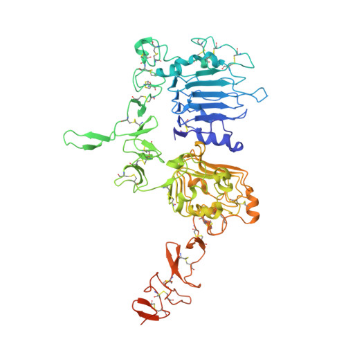
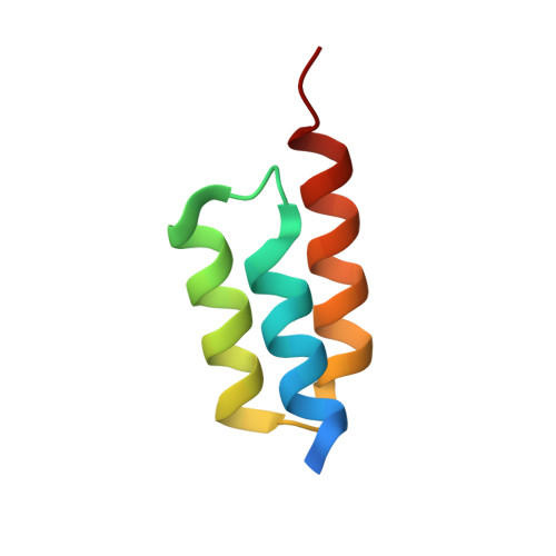
Structural basis for high-affinity HER2 receptor binding by an engineered protein.

Eigenbrot, C.Ultsch, M.Dubnovitsky, A.Abrahmsen, L.Hard, T.

(2010) Proc Natl Acad Sci U S A 107: 15039-15044

  • DOI: https://doi.org/10.1073/pnas.1005025107
  • Primary Citation of Related Structures:  
    2KZI, 2KZJ, 3MZW

  • PubMed Abstract: 

    The human epidermal growth factor receptor 2 (HER2) is specifically overexpressed in tumors of several cancers, including an aggressive form of breast cancer. It is therefore a target for both cancer diagnostics and therapy. The 58 amino acid residue Zher2 affibody molecule was previously engineered as a high-affinity binder of HER2. Here we determined the structure of Zher2 in solution and the crystal structure of Zher2 in complex with the HER2 extracellular domain. Zher2 binds to a conformational epitope on HER2 that is distant from those recognized by the therapeutic antibodies trastuzumab and pertuzumab. Its small size and lack of interference may provide Zher2 with advantages for diagnostic use or even for delivery of therapeutic agents to HER2-expressing tumors when trastuzumab or pertuzumab are already employed. Biophysical characterization shows that Zher2 is thermodynamically stable in the folded state yet undergoing conformational interconversion on a submillisecond time scale. The data suggest that it is the HER2-binding conformation that is formed transiently prior to binding. Still, binding is very strong with a dissociation constant K(D) = 22 pM, and perfect conformational homogeneity is therefore not necessarily required in engineered binding proteins. A comparison of the original Z domain scaffold to free and bound Zher2 structures reveals how high-affinity binding has evolved during selection and affinity maturation and suggests how a compromise between binding surface optimization and stability and dynamics of the unbound state has been reached.


  • Organizational Affiliation

    Department of Structural Biology, Genentech Inc, 1 DNA Way, South San Francisco, CA 94080, USA. eigenbrot.c@gene.com


Macromolecules
Find similar proteins by:  (by identity cutoff)  |  3D Structure
Entity ID: 1
MoleculeChains Sequence LengthOrganismDetailsImage
Tyrosine kinase-type cell surface receptor HER2624Homo sapiensMutation(s): 0 
Gene Names: ERBB2HER2MLN19NEUNGL
EC: 2.7.10.1
UniProt & NIH Common Fund Data Resources
Find proteins for P04626 (Homo sapiens)
Explore P04626 
Go to UniProtKB:  P04626
PHAROS:  P04626
GTEx:  ENSG00000141736 
Entity Groups  
Sequence Clusters30% Identity50% Identity70% Identity90% Identity95% Identity100% Identity
UniProt GroupP04626
Sequence Annotations
Expand
  • Reference Sequence
Find similar proteins by:  (by identity cutoff)  |  3D Structure
Entity ID: 2
MoleculeChains Sequence LengthOrganismDetailsImage
Immunoglobulin G-binding protein A58Staphylococcus aureusMutation(s): 14 
UniProt
Find proteins for P38507 (Staphylococcus aureus)
Explore P38507 
Go to UniProtKB:  P38507
Entity Groups  
Sequence Clusters30% Identity50% Identity70% Identity90% Identity95% Identity100% Identity
UniProt GroupP38507
Sequence Annotations
Expand
  • Reference Sequence
Oligosaccharides

Help

Entity ID: 3
MoleculeChains Length2D Diagram Glycosylation3D Interactions
2-acetamido-2-deoxy-beta-D-glucopyranose-(1-4)-2-acetamido-2-deoxy-beta-D-glucopyranose
C
2N-Glycosylation
Glycosylation Resources
GlyTouCan:  G42666HT
GlyCosmos:  G42666HT
GlyGen:  G42666HT
Small Molecules
Ligands 1 Unique
IDChains Name / Formula / InChI Key2D Diagram3D Interactions
NAG
Query on NAG

Download Ideal Coordinates CCD File 
D [auth A],
E [auth A]
2-acetamido-2-deoxy-beta-D-glucopyranose
C8 H15 N O6
OVRNDRQMDRJTHS-FMDGEEDCSA-N
Experimental Data & Validation

Experimental Data

  • Method: X-RAY DIFFRACTION
  • Resolution: 2.90 Å
  • R-Value Free: 0.278 
  • R-Value Work: 0.204 
  • R-Value Observed: 0.208 
  • Space Group: P 21 21 21
Unit Cell:
Length ( Å )Angle ( ˚ )
a = 71.726α = 90
b = 97.841β = 90
c = 148.283γ = 90
Software Package:
Software NamePurpose
PHASERphasing
REFMACrefinement
XDSdata reduction
XDSdata scaling

Structure Validation

View Full Validation Report


Ligand Structure Quality Assessment 


Entry History 

Deposition Data

Revision History  (Full details and data files)

  • Version 1.0: 2010-07-28
    Type: Initial release
  • Version 1.1: 2011-07-13
    Changes: Version format compliance
  • Version 1.2: 2017-11-08
    Changes: Derived calculations, Refinement description
  • Version 2.0: 2020-07-29
    Type: Remediation
    Reason: Carbohydrate remediation
    Changes: Atomic model, Data collection, Database references, Derived calculations, Structure summary
  • Version 2.1: 2023-09-06
    Changes: Data collection, Database references, Refinement description, Structure summary