3MYP

Crystal structure of tagatose-1,6-bisphosphate aldolase from Staphylococcus aureus


Experimental Data Snapshot

  • Method: X-RAY DIFFRACTION
  • Resolution: 2.99 Å
  • R-Value Free: 0.241 
  • R-Value Work: 0.203 
  • R-Value Observed: 0.205 

wwPDB Validation   3D Report Full Report


This is version 1.1 of the entry. See complete history


Literature

Crystal structures of LacD from Staphylococcus aureus and LacD.1 from Streptococcus pyogenes: Insights into substrate specificity and virulence gene regulation

Lee, S.J.Kim, H.S.Kim, D.J.Yoon, H.J.Kim, K.H.Yoon, J.Y.Suh, S.W.

(2011) FEBS Lett 585: 307-312

  • DOI: https://doi.org/10.1016/j.febslet.2010.12.038
  • Primary Citation of Related Structures:  
    3MYO, 3MYP

  • PubMed Abstract: 

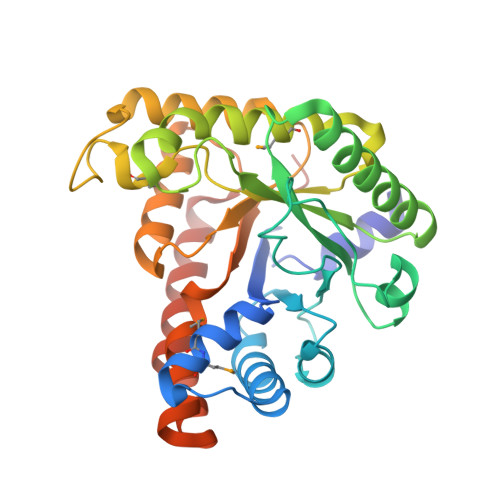
    Staphylococcus aureus LacD, a Class I tagatose-1,6-bisphosphate (TBP) aldolase, shows broadened substrate specificity by catalyzing the cleavage of 1,6-bisphosphate derivatives of D-tagatose, D-fructose, D-sorbose, and D-psicose. LacD.1 and LacD.2 are two closely-related Class I TBP aldolases in Streptococcus pyogenes. Here we have determined the crystal structures of S. aureus LacD and S. pyogenes LacD.1. Monomers of both enzymes are folded into a (β/α)(8) barrel and two monomers associate tightly to form a dimer in the crystals. The structures suggest that the residues E189 and S300 of rabbit muscle Class I fructose-1,6-bisphosphate (FBP) aldolase are important for substrate specificity. When we mutated the corresponding residues of S. aureus LacD, the mutants (L165E, L275S, and L165E/L275S) showed enhanced substrate specificity toward FBP.


  • Organizational Affiliation

    Department of Chemistry, College of Natural Sciences, Seoul National University, Seoul, Republic of Korea.


Macromolecules
Find similar proteins by:  (by identity cutoff)  |  3D Structure
Entity ID: 1
MoleculeChains Sequence LengthOrganismDetailsImage
Tagatose 1,6-diphosphate aldolase
A, B, C, D
334Staphylococcus aureus subsp. aureus COLMutation(s): 0 
Gene Names: lacDSACOL2183
EC: 4.1.2.40
UniProt
Find proteins for Q5HE13 (Staphylococcus aureus (strain COL))
Explore Q5HE13 
Go to UniProtKB:  Q5HE13
Entity Groups  
Sequence Clusters30% Identity50% Identity70% Identity90% Identity95% Identity100% Identity
UniProt GroupQ5HE13
Sequence Annotations
Expand
  • Reference Sequence
Small Molecules
Modified Residues  1 Unique
IDChains TypeFormula2D DiagramParent
MSE
Query on MSE
A, B, C, D
L-PEPTIDE LINKINGC5 H11 N O2 SeMET
Experimental Data & Validation

Experimental Data

  • Method: X-RAY DIFFRACTION
  • Resolution: 2.99 Å
  • R-Value Free: 0.241 
  • R-Value Work: 0.203 
  • R-Value Observed: 0.205 
  • Space Group: P 31
Unit Cell:
Length ( Å )Angle ( ˚ )
a = 104.899α = 90
b = 104.899β = 90
c = 115.863γ = 120
Software Package:
Software NamePurpose
HKL-2000data collection
SOLVEphasing
REFMACrefinement
HKL-2000data reduction
HKL-2000data scaling

Structure Validation

View Full Validation Report


Entry History 

Deposition Data

Revision History  (Full details and data files)

  • Version 1.0: 2011-01-26
    Type: Initial release
  • Version 1.1: 2011-07-13
    Changes: Version format compliance