3MMK

The structural basis for partial redundancy in a class of transcription factors, the lim-homeodomain proteins, in neural cell type specification


Experimental Data Snapshot

  • Method: X-RAY DIFFRACTION
  • Resolution: 2.16 Å
  • R-Value Free: 0.265 
  • R-Value Work: 0.235 
  • R-Value Observed: 0.237 

wwPDB Validation   3D Report Full Report


This is version 1.2 of the entry. See complete history


Literature

The structural basis for partial redundancy in a class of transcription factors, the lim-homeodomain proteins, in neural cell type specification.

Gadd, M.S.Bhati, M.Jeffries, C.M.Langley, D.B.Trewhella, J.Guss, J.M.Matthews, J.M.

(2011) J Biol Chem 

  • DOI: https://doi.org/10.1074/jbc.M111.248559
  • Primary Citation of Related Structures:  
    3MMK

  • PubMed Abstract: 

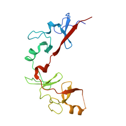
    Combinations of LIM homeodomain proteins form a transcriptional "LIM code" to direct the specification of neural cell types. Two paralogous pairs of LIM homeodomain proteins, LIM homeobox protein 3/4 (Lhx3/Lhx4) and Islet-1/2 (Isl1/Isl2), are expressed in developing ventral motor neurons. Lhx3 and Isl1 interact within a well characterized transcriptional complex that triggers motor neuron development, but it was not known whether Lhx4 and Isl2 could participate in equivalent complexes. We have identified an Lhx3-binding domain (LBD) in Isl2 based on sequence homology with the Isl1(LBD) and show that both Isl2(LBD) and Isl1(LBD) can bind each of Lhx3 and Lhx4. X-ray crystal- and small-angle x-ray scattering-derived solution structures of an Lhx4·Isl2 complex exhibit many similarities with that of Lhx3·Isl1; however, structural differences supported by mutagenic studies reveal differences in the mechanisms of binding. Differences in binding have implications for the mode of exchange of protein partners in transcriptional complexes and indicate a divergence in functions of Lhx3/4 and Isl1/2. The formation of weaker Lhx·Isl complexes would likely be masked by the availability of the other Lhx·Isl complexes in postmitotic motor neurons.


  • Organizational Affiliation

    School of Molecular Bioscience, The University of Sydney, New South Wales 2006, Australia.


Macromolecules
Find similar proteins by:  (by identity cutoff)  |  3D Structure
Entity ID: 1
MoleculeChains Sequence LengthOrganismDetailsImage
Fusion of LIM/homeobox protein Lhx4, linker, Insulin gene enhancer protein ISL-2
A, B
169Mus musculusMutation(s): 0 
Gene Names: Gsh-4Gsh4Isl2Lhx4
UniProt
Find proteins for Q9CXV0 (Mus musculus)
Explore Q9CXV0 
Go to UniProtKB:  Q9CXV0
Find proteins for P53776 (Mus musculus)
Explore P53776 
Go to UniProtKB:  P53776
Entity Groups  
Sequence Clusters30% Identity50% Identity70% Identity90% Identity95% Identity100% Identity
UniProt GroupsQ9CXV0P53776
Sequence Annotations
Expand
  • Reference Sequence
Small Molecules
Ligands 2 Unique
IDChains Name / Formula / InChI Key2D Diagram3D Interactions
ZN
Query on ZN

Download Ideal Coordinates CCD File 
C [auth A]
D [auth A]
E [auth A]
F [auth A]
J [auth B]
C [auth A],
D [auth A],
E [auth A],
F [auth A],
J [auth B],
K [auth B],
L [auth B],
M [auth B]
ZINC ION
Zn
PTFCDOFLOPIGGS-UHFFFAOYSA-N
CL
Query on CL

Download Ideal Coordinates CCD File 
G [auth A],
H [auth A],
I [auth A],
N [auth B]
CHLORIDE ION
Cl
VEXZGXHMUGYJMC-UHFFFAOYSA-M
Experimental Data & Validation

Experimental Data

  • Method: X-RAY DIFFRACTION
  • Resolution: 2.16 Å
  • R-Value Free: 0.265 
  • R-Value Work: 0.235 
  • R-Value Observed: 0.237 
  • Space Group: P 1 21 1
Unit Cell:
Length ( Å )Angle ( ˚ )
a = 46.823α = 90
b = 88.734β = 111.85
c = 49.851γ = 90
Software Package:
Software NamePurpose
ADSCdata collection
SOLVEphasing
PHENIXrefinement
HKL-2000data reduction
HKL-2000data scaling

Structure Validation

View Full Validation Report


Entry History 

Deposition Data

Revision History  (Full details and data files)

  • Version 1.0: 2011-07-13
    Type: Initial release
  • Version 1.1: 2011-11-16
    Changes: Database references, Structure summary
  • Version 1.2: 2017-06-28
    Changes: Source and taxonomy