3MK4

X-Ray structure of human PEX3 in complex with a PEX19 derived peptide


Experimental Data Snapshot

  • Method: X-RAY DIFFRACTION
  • Resolution: 2.42 Å
  • R-Value Free: 0.234 
  • R-Value Work: 0.194 
  • R-Value Observed: 0.196 

wwPDB Validation   3D Report Full Report


This is version 1.3 of the entry. See complete history


Literature

Insights into peroxisome function from the structure of PEX3 in complex with a soluble fragment of PEX19

Schmidt, F.Treiber, N.Zocher, G.Bjelic, S.Steinmetz, M.O.Kalbacher, H.Stehle, T.Dodt, G.

(2010) J Biol Chem 285: 25410-25417

  • DOI: https://doi.org/10.1074/jbc.M110.138503
  • Primary Citation of Related Structures:  
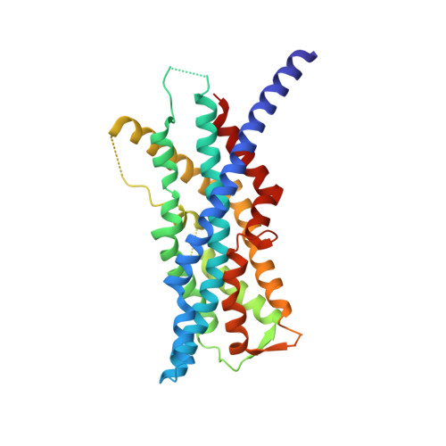
    3MK4

  • PubMed Abstract: 

    The human peroxins PEX3 and PEX19 play a central role in peroxisomal membrane biogenesis. The membrane-anchored PEX3 serves as the receptor for cytosolic PEX19, which in turn recognizes newly synthesized peroxisomal membrane proteins. After delivering these proteins to the peroxisomal membrane, PEX19 is recycled to the cytosol. The molecular mechanisms underlying these processes are not well understood. Here, we report the crystal structure of the cytosolic domain of PEX3 in complex with a PEX19-derived peptide. PEX3 adopts a novel fold that is best described as a large helical bundle. A hydrophobic groove at the membrane-distal end of PEX3 engages the PEX19 peptide with nanomolar affinity. Mutagenesis experiments identify phenylalanine 29 in PEX19 as critical for this interaction. Because key PEX3 residues involved in complex formation are highly conserved across species, the observed binding mechanism is of general biological relevance.


  • Organizational Affiliation

    Interfaculty Institute for Biochemistry, University of Tübingen, 72076 Tübingen, Germany.


Macromolecules
Find similar proteins by:  (by identity cutoff)  |  3D Structure
Entity ID: 1
MoleculeChains Sequence LengthOrganismDetailsImage
Peroxisomal biogenesis factor 3334Homo sapiensMutation(s): 1 
Gene Names: PEX3
UniProt & NIH Common Fund Data Resources
Find proteins for P56589 (Homo sapiens)
Explore P56589 
Go to UniProtKB:  P56589
PHAROS:  P56589
GTEx:  ENSG00000034693 
Entity Groups  
Sequence Clusters30% Identity50% Identity70% Identity90% Identity95% Identity100% Identity
UniProt GroupP56589
Sequence Annotations
Expand
  • Reference Sequence

Find similar proteins by:  Sequence   |   3D Structure  

Entity ID: 2
MoleculeChains Sequence LengthOrganismDetailsImage
Peroxisomal biogenesis factor 1920N/AMutation(s): 0 
UniProt & NIH Common Fund Data Resources
Find proteins for P40855 (Homo sapiens)
Explore P40855 
Go to UniProtKB:  P40855
PHAROS:  P40855
GTEx:  ENSG00000162735 
Entity Groups  
Sequence Clusters30% Identity50% Identity70% Identity90% Identity95% Identity100% Identity
UniProt GroupP40855
Sequence Annotations
Expand
  • Reference Sequence
Experimental Data & Validation

Experimental Data

  • Method: X-RAY DIFFRACTION
  • Resolution: 2.42 Å
  • R-Value Free: 0.234 
  • R-Value Work: 0.194 
  • R-Value Observed: 0.196 
  • Space Group: P 1 21 1
Unit Cell:
Length ( Å )Angle ( ˚ )
a = 38.47α = 90
b = 65.67β = 91.53
c = 61.58γ = 90
Software Package:
Software NamePurpose
MxCuBEdata collection
PHASERphasing
REFMACrefinement
XDSdata reduction
XSCALEdata scaling

Structure Validation

View Full Validation Report


Entry History 

Deposition Data

Revision History  (Full details and data files)

  • Version 1.0: 2010-06-30
    Type: Initial release
  • Version 1.1: 2011-07-13
    Changes: Version format compliance
  • Version 1.2: 2013-10-16
    Changes: Derived calculations
  • Version 1.3: 2014-02-19
    Changes: Database references