


Diversity in the cross-beta spine structure of prion peptides
Lee, S., Yee, V.C.To be published.
Experimental Data Snapshot
wwPDB Validation 3D Report Full Report

Find similar proteins by: Sequence | 3D Structure
Entity ID: 1 | |||||
|---|---|---|---|---|---|
| Molecule | Chains | Sequence Length | Organism | Details | Image |
| Major prion protein | 6 | N/A | Mutation(s): 0 | 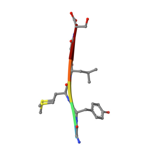 | |
UniProt & NIH Common Fund Data Resources | |||||
Find proteins for P04156 (Homo sapiens) Explore P04156 Go to UniProtKB: P04156 | |||||
PHAROS: P04156 GTEx: ENSG00000171867 | |||||
Entity Groups | |||||
| UniProt Group | P04156 | ||||
Sequence AnnotationsExpand | |||||
| |||||
| Length ( Å ) | Angle ( ˚ ) |
|---|---|
| a = 9.439 | α = 90 |
| b = 17.792 | β = 90 |
| c = 44.561 | γ = 90 |
| Software Name | Purpose |
|---|---|
| DENZO | data reduction |
| SCALEPACK | data scaling |
| REFMAC | refinement |
| PDB_EXTRACT | data extraction |
| HKL-2000 | data collection |
| HKL-2000 | data reduction |
| HKL-2000 | data scaling |
| SHELXD | phasing |
RCSB PDB (citation) is hosted by
RCSB PDB is a member of the

