3LU9

Crystal structure of human thrombin mutant S195A in complex with the extracellular fragment of human PAR1


Experimental Data Snapshot

  • Method: X-RAY DIFFRACTION
  • Resolution: 1.80 Å
  • R-Value Free: 0.236 
  • R-Value Work: 0.193 
  • R-Value Observed: 0.195 

wwPDB Validation   3D Report Full Report


Ligand Structure Quality Assessment 


This is version 1.4 of the entry. See complete history


Literature

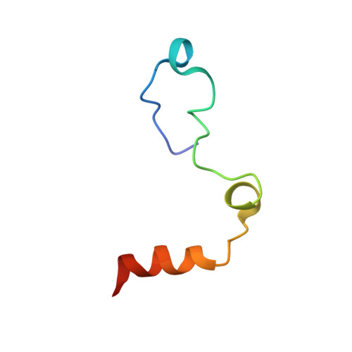
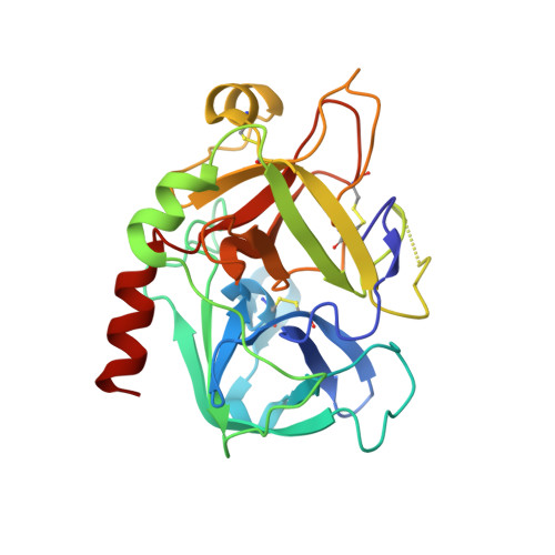
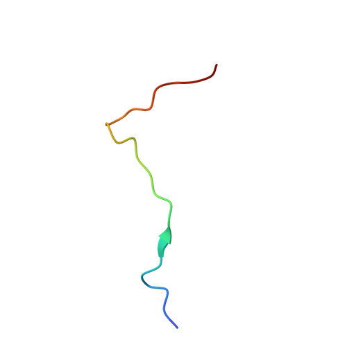
Crystal structure of thrombin bound to the uncleaved extracellular fragment of PAR1.

Gandhi, P.S.Chen, Z.Di Cera, E.

(2010) J Biol Chem 285: 15393-15398

  • DOI: https://doi.org/10.1074/jbc.M110.115337
  • Primary Citation of Related Structures:  
    3LU9

  • PubMed Abstract: 

    Abundant structural information exists on how thrombin recognizes ligands at the active site or at exosites separate from the active site region, but remarkably little is known about how thrombin recognizes substrates that bridge both the active site and exosite I. The case of the protease-activated receptor PAR1 is particularly relevant in view of the plethora of biological effects associated with its activation by thrombin. Here, we present the 1.8 A resolution structure of thrombin S195A in complex with a 30-residue long uncleaved extracellular fragment of PAR1 that documents for the first time a productive binding mode bridging the active site and exosite I. The structure reveals two unexpected features of the thrombin-PAR1 interaction. The acidic P3 residue of PAR1, Asp(39), does not hinder binding to the active site and actually makes favorable interactions with Gly(219) of thrombin. The tethered ligand domain shows a considerable degree of disorder even when bound to thrombin. The results fill a significant gap in our understanding of the molecular mechanisms of recognition by thrombin in ways that are relevant to other physiological substrates.


  • Organizational Affiliation

    Department of Biochemistry and Molecular Biology, Saint Louis University School of Medicine, St. Louis, Missouri 63104.


Macromolecules
Find similar proteins by:  (by identity cutoff)  |  3D Structure
Entity ID: 1
MoleculeChains Sequence LengthOrganismDetailsImage
Prothrombin
A, D
46Homo sapiensMutation(s): 0 
Gene Names: F2Human
EC: 3.4.21.5
UniProt & NIH Common Fund Data Resources
Find proteins for P00734 (Homo sapiens)
Explore P00734 
Go to UniProtKB:  P00734
PHAROS:  P00734
GTEx:  ENSG00000180210 
Entity Groups  
Sequence Clusters30% Identity50% Identity70% Identity90% Identity95% Identity100% Identity
UniProt GroupP00734
Sequence Annotations
Expand
  • Reference Sequence
Find similar proteins by:  (by identity cutoff)  |  3D Structure
Entity ID: 2
MoleculeChains Sequence LengthOrganismDetailsImage
Prothrombin
B, E
259Homo sapiensMutation(s): 1 
Gene Names: F2
EC: 3.4.21.5
UniProt & NIH Common Fund Data Resources
Find proteins for P00734 (Homo sapiens)
Explore P00734 
Go to UniProtKB:  P00734
PHAROS:  P00734
GTEx:  ENSG00000180210 
Entity Groups  
Sequence Clusters30% Identity50% Identity70% Identity90% Identity95% Identity100% Identity
UniProt GroupP00734
Sequence Annotations
Expand
  • Reference Sequence

Find similar proteins by:  Sequence   |   3D Structure  

Entity ID: 3
MoleculeChains Sequence LengthOrganismDetailsImage
Proteinase-activated receptor 1
C, F
25Homo sapiensMutation(s): 0 
Gene Names: F2RCF2RPAR1TR
UniProt & NIH Common Fund Data Resources
Find proteins for P25116 (Homo sapiens)
Explore P25116 
Go to UniProtKB:  P25116
PHAROS:  P25116
GTEx:  ENSG00000181104 
Entity Groups  
Sequence Clusters30% Identity50% Identity70% Identity90% Identity95% Identity100% Identity
UniProt GroupP25116
Sequence Annotations
Expand
  • Reference Sequence
Experimental Data & Validation

Experimental Data

  • Method: X-RAY DIFFRACTION
  • Resolution: 1.80 Å
  • R-Value Free: 0.236 
  • R-Value Work: 0.193 
  • R-Value Observed: 0.195 
  • Space Group: P 1
Unit Cell:
Length ( Å )Angle ( ˚ )
a = 46.334α = 76.39
b = 50.171β = 83.93
c = 85.502γ = 73.74
Software Package:
Software NamePurpose
ADSCdata collection
MOLREPphasing
REFMACrefinement
HKL-2000data reduction
HKL-2000data scaling

Structure Validation

View Full Validation Report


Ligand Structure Quality Assessment 


Entry History 

Deposition Data

Revision History  (Full details and data files)

  • Version 1.0: 2010-03-16
    Type: Initial release
  • Version 1.1: 2011-07-13
    Changes: Version format compliance
  • Version 1.2: 2020-07-29
    Type: Remediation
    Reason: Carbohydrate remediation
    Changes: Advisory, Data collection, Derived calculations, Structure summary
  • Version 1.3: 2021-10-13
    Changes: Advisory, Database references, Structure summary
  • Version 1.4: 2023-09-06
    Changes: Data collection, Refinement description