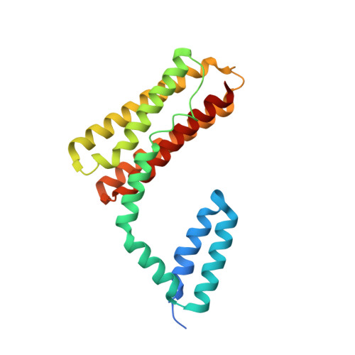
Crystal structure of CCM3, a cerebral cavernous malformation protein critical for vascular integrity.
Li, X., Zhang, R., Zhang, H., He, Y., Ji, W., Min, W., Boggon, T.J.(2010) J Biol Chem 285: 24099-24107
- PubMed: 20489202
- DOI: https://doi.org/10.1074/jbc.M110.128470
- Primary Citation of Related Structures:
3L8I, 3L8J - PubMed Abstract:
CCM3 mutations are associated with cerebral cavernous malformation (CCM), a disease affecting 0.1-0.5% of the human population. CCM3 (PDCD10, TFAR15) is thought to form a CCM complex with CCM1 and CCM2; however, the molecular basis for these interactions is not known. We have determined the 2.5 A crystal structure of CCM3. This structure shows an all alpha-helical protein containing two domains, an N-terminal dimerization domain with a fold not previously observed, and a C-terminal focal adhesion targeting (FAT)-homology domain. We show that CCM3 binds CCM2 via this FAT-homology domain and that mutation of a highly conserved FAK-like hydrophobic pocket (HP1) abrogates CCM3-CCM2 interaction. This CCM3 FAT-homology domain also interacts with paxillin LD motifs using the same surface, and partial CCM3 co-localization with paxillin in cells is lost on HP1 mutation. Disease-related CCM3 truncations affect the FAT-homology domain suggesting a role for the FAT-homology domain in the etiology of CCM.
Organizational Affiliation:
Department of Pharmacology, Yale University School of Medicine, New Haven, CT 06520, USA.