3KPZ

Crystal structure of a novel vitamin D3 analogue, ZK203278 showing dissociated profile


Experimental Data Snapshot

  • Method: X-RAY DIFFRACTION
  • Resolution: 1.90 Å
  • R-Value Free: 0.228 
  • R-Value Work: 0.195 
  • R-Value Observed: 0.228 

wwPDB Validation   3D Report Full Report


Ligand Structure Quality Assessment 


This is version 1.3 of the entry. See complete history


Literature

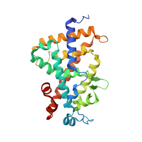
Crystal structure of a vitamin D3 analog, ZK203278, showing dissociated profile.

Rochel, N.Moras, D.

(2012) Anticancer Res 32: 335-339

  • Primary Citation of Related Structures:  
    3KPZ

  • PubMed Abstract: 

    The plethora of actions of 1α,25-dihydroxyvitamin D(3), the active form of the seco-steroid hormone vitamin D, in various systems suggested wide clinical applications in treatments for renal osteodystrophy, osteoporosis, psoriasis, cancer, autoimmune diseases and prevention of graft rejection. However, the major side-effects of hypercalcemia of VDR ligands limit their use. ZK203278, a novel synthetic analog has been shown to act as a potent immunomodulator and presents dissociated biologic profile with low calcemic side-effects. Here, we described the crystal structures of the hVDR ligand-binding domain in complex with ZK203278 and determined its correlation with its specific dissociated biologic profile. The VDR/ZK203278 structure, in comparison with VDR/1α,25-dihydroxyvitamin D(3), shows specific interactions of the thiazole group of ZK203278 with residues of H3, H11 and H12. These specific interactions may lead to altered selective interactions with co-regulators and consequently to the dissociated biologic profile of this novel ligand.


  • Organizational Affiliation

    Institut de Génétique et de Biologie Moléculaire et Cellulaire (IGBMC), Institut National de Santé et de Recherche Médicale (INSERM) U964/Centre National de Recherche Scientifique (CNRS) UMR 7104/Université de Strasbourg, 67404 Illkirch, France. rochel@igbmc.fr


Macromolecules
Find similar proteins by:  (by identity cutoff)  |  3D Structure
Entity ID: 1
MoleculeChains Sequence LengthOrganismDetailsImage
Vitamin D3 receptor259Homo sapiensMutation(s): 0 
Gene Names: VDRNR1I1
UniProt & NIH Common Fund Data Resources
Find proteins for P11473 (Homo sapiens)
Explore P11473 
Go to UniProtKB:  P11473
PHAROS:  P11473
GTEx:  ENSG00000111424 
Entity Groups  
Sequence Clusters30% Identity50% Identity70% Identity90% Identity95% Identity100% Identity
UniProt GroupP11473
Sequence Annotations
Expand
  • Reference Sequence
Small Molecules
Ligands 1 Unique
IDChains Name / Formula / InChI Key2D Diagram3D Interactions
ZNE
Query on ZNE

Download Ideal Coordinates CCD File 
B [auth A](1R,3S,5Z)-5-[(2E)-2-{(1R,3aS,7aR)-1-[(1R,5S)-5-hydroxy-1-methyl-5-(1,3-thiazol-2-yl)pentyl]-7a-methyloctahydro-4H-inden-4-ylidene}ethylidene]-4-methylidenecyclohexane-1,3-diol
C28 H41 N O3 S
AZVJHDPZXYTKQV-LCRUICLWSA-N
Experimental Data & Validation

Experimental Data

  • Method: X-RAY DIFFRACTION
  • Resolution: 1.90 Å
  • R-Value Free: 0.228 
  • R-Value Work: 0.195 
  • R-Value Observed: 0.228 
  • Space Group: P 21 21 21
Unit Cell:
Length ( Å )Angle ( ˚ )
a = 44.887α = 90
b = 51.892β = 90
c = 132.204γ = 90
Software Package:
Software NamePurpose
CNSrefinement
PDB_EXTRACTdata extraction
HKL-2000data reduction
HKL-2000data scaling
CNSphasing

Structure Validation

View Full Validation Report


Ligand Structure Quality Assessment 


Entry History 

Deposition Data

Revision History  (Full details and data files)

  • Version 1.0: 2011-10-12
    Type: Initial release
  • Version 1.1: 2011-10-26
    Changes: Structure summary
  • Version 1.2: 2012-04-25
    Changes: Database references
  • Version 1.3: 2023-09-06
    Changes: Data collection, Database references, Derived calculations, Refinement description