3K65

Crystal Structure of Prethombin-2/Fragment-2 Complex


Experimental Data Snapshot

  • Method: X-RAY DIFFRACTION
  • Resolution: 1.85 Å
  • R-Value Free: 0.229 
  • R-Value Work: 0.213 
  • R-Value Observed: 0.219 

wwPDB Validation   3D Report Full Report


This is version 1.5 of the entry. See complete history


Literature

Structural transitions during prothrombin activation: On the importance of fragment 2.

Adams, T.E.Huntington, J.A.

(2016) Biochimie 122: 235-242

  • DOI: https://doi.org/10.1016/j.biochi.2015.09.013
  • Primary Citation of Related Structures:  
    3K65

  • PubMed Abstract: 

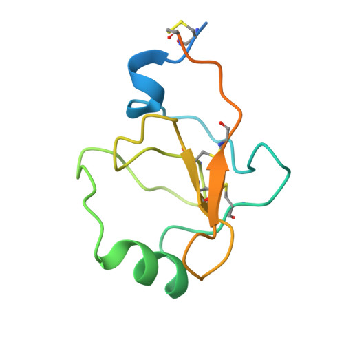
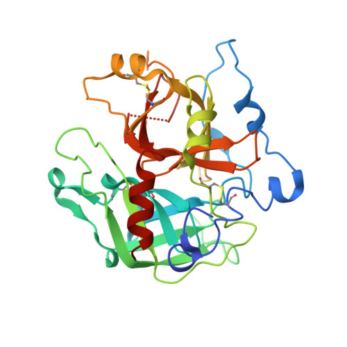
    Prothrombin is activated to thrombin by the prothrombinase complex through sequential cleavage at two distinct sites. This occurs at sites of vascular injury in a highly regulated cascade of serine protease and cofactor activation, where activated platelets provide a suitable surface for protease/cofactor/substrate assembly. The precise structural and conformational changes undergone during the transition from prothrombin to thrombin have been studied for decades, and several structures of prothrombin fragments along the activation pathway have been solved. Here we present a new structure analyzed in context of other recent structures and biochemical studies. What emerges is an unexpected mechanism that involves a change in the mode of binding of the F2 domain (fragment 2) on the catalytic domain after cleavage at Arg320, and a subsequent reorientation of the linker between the F2 and catalytic domain to present the Arg271 site for cleavage.


  • Organizational Affiliation

    Cambridge Institute for Medical Research, Department of Haematology, University of Cambridge, Wellcome Trust/MRC Building, Hills Road, Cambridge, CB2 0XY, United Kingdom.


Macromolecules
Find similar proteins by:  (by identity cutoff)  |  3D Structure
Entity ID: 1
MoleculeChains Sequence LengthOrganismDetailsImage
Prothrombin116Homo sapiensMutation(s): 0 
EC: 3.4.21.5
UniProt & NIH Common Fund Data Resources
Find proteins for P00734 (Homo sapiens)
Explore P00734 
Go to UniProtKB:  P00734
PHAROS:  P00734
GTEx:  ENSG00000180210 
Entity Groups  
Sequence Clusters30% Identity50% Identity70% Identity90% Identity95% Identity100% Identity
UniProt GroupP00734
Sequence Annotations
Expand
  • Reference Sequence
Find similar proteins by:  (by identity cutoff)  |  3D Structure
Entity ID: 2
MoleculeChains Sequence LengthOrganismDetailsImage
Prothrombin308Homo sapiensMutation(s): 1 
Gene Names: F2
EC: 3.4.21.5
UniProt & NIH Common Fund Data Resources
Find proteins for P00734 (Homo sapiens)
Explore P00734 
Go to UniProtKB:  P00734
PHAROS:  P00734
GTEx:  ENSG00000180210 
Entity Groups  
Sequence Clusters30% Identity50% Identity70% Identity90% Identity95% Identity100% Identity
UniProt GroupP00734
Sequence Annotations
Expand
  • Reference Sequence
Experimental Data & Validation

Experimental Data

  • Method: X-RAY DIFFRACTION
  • Resolution: 1.85 Å
  • R-Value Free: 0.229 
  • R-Value Work: 0.213 
  • R-Value Observed: 0.219 
  • Space Group: P 41 21 2
Unit Cell:
Length ( Å )Angle ( ˚ )
a = 73.53α = 90
b = 73.53β = 90
c = 205.88γ = 90
Software Package:
Software NamePurpose
MOSFLMdata reduction
SCALAdata scaling
PHASERphasing
CNSrefinement
PDB_EXTRACTdata extraction
DNAdata collection

Structure Validation

View Full Validation Report


Entry History 

Deposition Data

Revision History  (Full details and data files)

  • Version 1.0: 2010-09-29
    Type: Initial release
  • Version 1.1: 2011-07-13
    Changes: Version format compliance
  • Version 1.2: 2013-09-25
    Changes: Derived calculations
  • Version 1.3: 2017-07-05
    Changes: Database references
  • Version 1.4: 2021-10-13
    Changes: Database references, Derived calculations
  • Version 1.5: 2023-09-06
    Changes: Data collection, Refinement description