3J41

Pseudo-atomic model of the Aquaporin-0/Calmodulin complex derived from electron microscopy


Experimental Data Snapshot

  • Method: ELECTRON MICROSCOPY
  • Resolution: 25.0 Å
  • Aggregation State: PARTICLE 
  • Reconstruction Method: SINGLE PARTICLE 

wwPDB Validation   3D Report Full Report


This is version 1.4 of the entry. See complete history


Literature

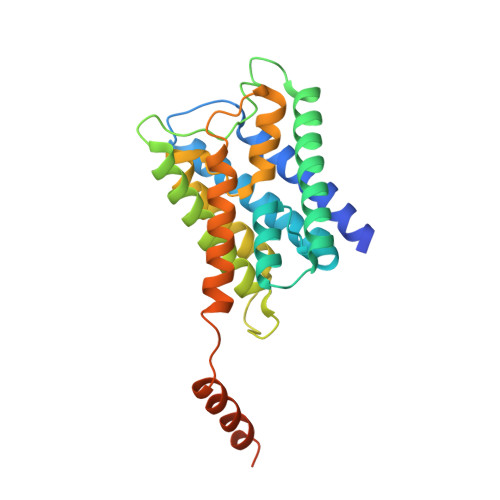
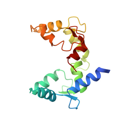
Allosteric mechanism of water-channel gating by Ca(2+)-calmodulin.

Reichow, S.L.Clemens, D.M.Freites, J.A.Nemeth-Cahalan, K.L.Heyden, M.Tobias, D.J.Hall, J.E.Gonen, T.

(2013) Nat Struct Mol Biol 20: 1085-1092

  • DOI: https://doi.org/10.1038/nsmb.2630
  • Primary Citation of Related Structures:  
    3J41

  • PubMed Abstract: 

    Calmodulin (CaM) is a universal regulatory protein that communicates the presence of calcium to its molecular targets and correspondingly modulates their function. This key signaling protein is important for controlling the activity of hundreds of membrane channels and transporters. However, understanding of the structural mechanisms driving CaM regulation of full-length membrane proteins has remained elusive. In this study, we determined the pseudoatomic structure of full-length mammalian aquaporin-0 (AQP0, Bos taurus) in complex with CaM, using EM to elucidate how this signaling protein modulates water-channel function. Molecular dynamics and functional mutation studies reveal how CaM binding inhibits AQP0 water permeability by allosterically closing the cytoplasmic gate of AQP0. Our mechanistic model provides new insight, only possible in the context of the fully assembled channel, into how CaM regulates multimeric channels by facilitating cooperativity between adjacent subunits.


  • Organizational Affiliation

    1] Howard Hughes Medical Institute, Janelia Farm Research Campus, Ashburn, Virginia, USA. [2].


Macromolecules
Find similar proteins by:  (by identity cutoff)  |  3D Structure
Entity ID: 1
MoleculeChains Sequence LengthOrganismDetailsImage
Lens fiber major intrinsic protein
A, B, C, D
263Ovis ariesMutation(s): 0 
Membrane Entity: Yes 
UniProt
Find proteins for Q6J8I9 (Ovis aries)
Explore Q6J8I9 
Go to UniProtKB:  Q6J8I9
Entity Groups  
Sequence Clusters30% Identity50% Identity70% Identity90% Identity95% Identity100% Identity
UniProt GroupQ6J8I9
Sequence Annotations
Expand
  • Reference Sequence
Find similar proteins by:  (by identity cutoff)  |  3D Structure
Entity ID: 2
MoleculeChains Sequence LengthOrganismDetailsImage
Calmodulin
E, F
149Homo sapiensMutation(s): 0 
Gene Names: 
CALM1CALMCAMCAM1CALM2CAM2CAMBCALM3CALML2CAM3...
CALM1CALMCAMCAM1CALM2CAM2CAMBCALM3CALML2CAM3CAMCCAMIII

UniProt & NIH Common Fund Data Resources
Find proteins for P0DP23 (Homo sapiens)
Explore P0DP23 
Go to UniProtKB:  P0DP23
PHAROS:  P0DP23
GTEx:  ENSG00000198668 
Entity Groups  
Sequence Clusters30% Identity50% Identity70% Identity90% Identity95% Identity100% Identity
UniProt GroupP0DP23
Sequence Annotations
Expand
  • Reference Sequence
Experimental Data & Validation

Experimental Data

  • Method: ELECTRON MICROSCOPY
  • Resolution: 25.0 Å
  • Aggregation State: PARTICLE 
  • Reconstruction Method: SINGLE PARTICLE 
EM Software:
TaskSoftware PackageVersion
RECONSTRUCTIONFREALIGN
RECONSTRUCTIONSPIDER

Structure Validation

View Full Validation Report


Entry History 

Deposition Data

Revision History  (Full details and data files)

  • Version 1.0: 2013-07-31
    Type: Initial release
  • Version 1.1: 2013-08-14
    Changes: Database references
  • Version 1.2: 2013-09-18
    Changes: Database references
  • Version 1.3: 2018-07-18
    Changes: Data collection
  • Version 1.4: 2024-02-21
    Changes: Data collection, Database references, Derived calculations, Refinement description