3IPQ

X-ray structure of GW3965 synthetic agonist bound to the LXR-alpha


Experimental Data Snapshot

  • Method: X-RAY DIFFRACTION
  • Resolution: 2.00 Å
  • R-Value Free: 0.234 
  • R-Value Work: 0.199 
  • R-Value Observed: 0.201 

wwPDB Validation   3D Report Full Report


Ligand Structure Quality Assessment 


This is version 1.2 of the entry. See complete history


Literature

X-ray structures of the LXRalpha LBD in its homodimeric form and implications for heterodimer signaling.

Fradera, X.Vu, D.Nimz, O.Skene, R.Hosfield, D.Wynands, R.Cooke, A.J.Haunso, A.King, A.Bennett, D.J.McGuire, R.Uitdehaag, J.C.

(2010) J Mol Biol 399: 120-132

  • DOI: https://doi.org/10.1016/j.jmb.2010.04.005
  • Primary Citation of Related Structures:  
    3IPQ, 3IPS, 3IPU

  • PubMed Abstract: 

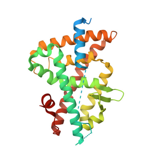
    Liver X receptors (LXRs) are nuclear receptors that are central regulators of cholesterol homeostasis, and synthetic LXR agonists have shown promise as promoters of reverse cholesterol transport and anti-inflammatory agents. Here, we present three X-ray structures of three different agonists bound to the ligand binding domain of LXRalpha. These compounds are GW3965, F(3)methylAA, and a benzisoxazole urea, and we show that these diverse chemical scaffolds address common structural themes, leading to high binding affinity for LXR. Our structures show the LXRalpha ligand binding domain in its homodimeric form, an arrangement previously thought to be stereochemically difficult. A comparison with existing structures of the LXRbeta homodimer and LXRalpha:RXR (retinoid X receptor) heterodimers explains differences in dimer affinity and leads us to propose a model for allosteric activation in nuclear receptor dimers, in which an unactivated RXR partner provides an inhibitory tail wrap to the cofactor binding pocket of LXR.


  • Organizational Affiliation

    Department of Chemistry, Merck Research Labs, Newhouse, ML1 5SH, Scotland, UK.


Macromolecules
Find similar proteins by:  (by identity cutoff)  |  3D Structure
Entity ID: 1
MoleculeChains Sequence LengthOrganismDetailsImage
Oxysterols receptor LXR-alpha283Homo sapiensMutation(s): 0 
Gene Names: LXRANR1H3
UniProt & NIH Common Fund Data Resources
Find proteins for Q13133 (Homo sapiens)
Explore Q13133 
Go to UniProtKB:  Q13133
PHAROS:  Q13133
GTEx:  ENSG00000025434 
Entity Groups  
Sequence Clusters30% Identity50% Identity70% Identity90% Identity95% Identity100% Identity
UniProt GroupQ13133
Sequence Annotations
Expand
  • Reference Sequence

Find similar proteins by:  Sequence   |   3D Structure  

Entity ID: 2
MoleculeChains Sequence LengthOrganismDetailsImage
Nuclear receptor coactivator 125N/AMutation(s): 0 
EC: 2.3.1.48
UniProt & NIH Common Fund Data Resources
Find proteins for Q15788 (Homo sapiens)
Explore Q15788 
Go to UniProtKB:  Q15788
PHAROS:  Q15788
GTEx:  ENSG00000084676 
Entity Groups  
Sequence Clusters30% Identity50% Identity70% Identity90% Identity95% Identity100% Identity
UniProt GroupQ15788
Sequence Annotations
Expand
  • Reference Sequence
Small Molecules
Ligands 2 Unique
IDChains Name / Formula / InChI Key2D Diagram3D Interactions
965
Query on 965

Download Ideal Coordinates CCD File 
C [auth A][3-(3-{[2-chloro-3-(trifluoromethyl)benzyl](2,2-diphenylethyl)amino}propoxy)phenyl]acetic acid
C33 H31 Cl F3 N O3
NAXSRXHZFIBFMI-UHFFFAOYSA-N
SO4
Query on SO4

Download Ideal Coordinates CCD File 
D [auth B]SULFATE ION
O4 S
QAOWNCQODCNURD-UHFFFAOYSA-L
Binding Affinity Annotations 
IDSourceBinding Affinity
965 Binding MOAD:  3IPQ Ki: 27 (nM) from 1 assay(s)
PDBBind:  3IPQ Ki: 27 (nM) from 1 assay(s)
BindingDB:  3IPQ IC50: min: 100, max: 235 (nM) from 3 assay(s)
EC50: min: 22, max: 1300 (nM) from 13 assay(s)
Experimental Data & Validation

Experimental Data

  • Method: X-RAY DIFFRACTION
  • Resolution: 2.00 Å
  • R-Value Free: 0.234 
  • R-Value Work: 0.199 
  • R-Value Observed: 0.201 
  • Space Group: I 4 2 2
Unit Cell:
Length ( Å )Angle ( ˚ )
a = 125.593α = 90
b = 125.593β = 90
c = 92.405γ = 90
Software Package:
Software NamePurpose
ADSCdata collection
AMoREphasing
REFMACrefinement
MOSFLMdata reduction
SCALAdata scaling

Structure Validation

View Full Validation Report


Ligand Structure Quality Assessment 


Entry History 

Deposition Data

Revision History  (Full details and data files)

  • Version 1.0: 2010-06-02
    Type: Initial release
  • Version 1.1: 2011-07-13
    Changes: Version format compliance
  • Version 1.2: 2024-02-21
    Changes: Data collection, Database references, Derived calculations