3HV4

Human p38 MAP Kinase in Complex with RL51


Experimental Data Snapshot

  • Method: X-RAY DIFFRACTION
  • Resolution: 2.60 Å
  • R-Value Free: 0.271 
  • R-Value Work: 0.194 
  • R-Value Observed: 0.197 

wwPDB Validation   3D Report Full Report


Ligand Structure Quality Assessment 


This is version 1.3 of the entry. See complete history


Literature

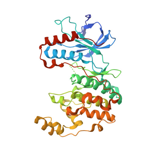
Displacement assay for the detection of stabilizers of inactive kinase conformations.

Kluter, S.Grutter, C.Naqvi, T.Rabiller, M.Simard, J.R.Pawar, V.Getlik, M.Rauh, D.

(2010) J Med Chem 53: 357-367

  • DOI: https://doi.org/10.1021/jm901297e
  • Primary Citation of Related Structures:  
    3HV3, 3HV4, 3HV5, 3HV6, 3HV7

  • PubMed Abstract: 

    Targeting protein kinases with small molecules outside the highly conserved ATP pocket to stabilize inactive kinase conformations is becoming a more desirable approach in kinase inhibitor research, since these molecules have advanced pharmacological properties compared to compounds exclusively targeting the ATP pocket. Traditional screening approaches for kinase inhibitors are often based on enzyme activity, but they may miss inhibitors that stabilize inactive kinase conformations by enriching the active state of the kinase. Here we present the development of a kinase binding assay employing a pyrazolourea type III inhibitor and enzyme fragment complementation (EFC) technology that is suitable to screen stabilizers of enzymatically inactive kinases. To validate this assay system, we report the binding characteristics of a series of kinase inhibitors to inactive p38alpha and JNK2. Additionally, we present protein X-ray crystallography studies to examine the binding modes of potent quinoline-based DFG-out binders in p38alpha.


  • Organizational Affiliation

    Chemical Genomics Centre of the Max Planck Society, Otto-Hahn-Strasse 15, D-44227 Dortmund, Germany.


Macromolecules
Find similar proteins by:  (by identity cutoff)  |  3D Structure
Entity ID: 1
MoleculeChains Sequence LengthOrganismDetailsImage
Mitogen-activated protein kinase 14
A, B
360Homo sapiensMutation(s): 0 
Gene Names: MAPK14CSBPCSBP1CSBP2CSPB1MXI2
EC: 2.7.11.24
UniProt & NIH Common Fund Data Resources
Find proteins for Q16539 (Homo sapiens)
Explore Q16539 
Go to UniProtKB:  Q16539
PHAROS:  Q16539
GTEx:  ENSG00000112062 
Entity Groups  
Sequence Clusters30% Identity50% Identity70% Identity90% Identity95% Identity100% Identity
UniProt GroupQ16539
Sequence Annotations
Expand
  • Reference Sequence
Small Molecules
Binding Affinity Annotations 
IDSourceBinding Affinity
L51 BindingDB:  3HV4 IC50: min: 290, max: 310 (nM) from 2 assay(s)
PDBBind:  3HV4 IC50: 310 (nM) from 1 assay(s)
Experimental Data & Validation

Experimental Data

  • Method: X-RAY DIFFRACTION
  • Resolution: 2.60 Å
  • R-Value Free: 0.271 
  • R-Value Work: 0.194 
  • R-Value Observed: 0.197 
  • Space Group: P 1 21 1
Unit Cell:
Length ( Å )Angle ( ˚ )
a = 70.64α = 90
b = 74.06β = 93.9
c = 72γ = 90
Software Package:
Software NamePurpose
XSCALEdata scaling
PHASERphasing
REFMACrefinement
PDB_EXTRACTdata extraction
XDSdata scaling

Structure Validation

View Full Validation Report


Ligand Structure Quality Assessment 


Entry History 

Deposition Data

Revision History  (Full details and data files)

  • Version 1.0: 2009-11-17
    Type: Initial release
  • Version 1.1: 2011-07-13
    Changes: Advisory, Version format compliance
  • Version 1.2: 2020-07-29
    Type: Remediation
    Reason: Carbohydrate remediation
    Changes: Data collection, Database references, Derived calculations, Structure summary
  • Version 1.3: 2023-09-06
    Changes: Data collection, Database references, Refinement description, Structure summary