3HU3

Structure of p97 N-D1 R155H mutant in complex with ATPgS


Experimental Data Snapshot

  • Method: X-RAY DIFFRACTION
  • Resolution: 2.20 Å
  • R-Value Free: 0.193 
  • R-Value Work: 0.171 
  • R-Value Observed: 0.172 

wwPDB Validation   3D Report Full Report


This is version 1.3 of the entry. See complete history


Literature

A novel ATP-dependent conformation in p97 N-D1 fragment revealed by crystal structures of disease-related mutants.

Tang, W.K.Li, D.Li, C.C.Esser, L.Dai, R.Guo, L.Xia, D.

(2010) EMBO J 29: 2217-2229

  • DOI: https://doi.org/10.1038/emboj.2010.104
  • Primary Citation of Related Structures:  
    3HU1, 3HU2, 3HU3

  • PubMed Abstract: 

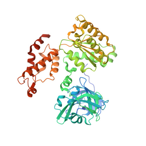
    Mutations in p97, a major cytosolic AAA (ATPases associated with a variety of cellular activities) chaperone, cause inclusion body myopathy associated with Paget's disease of the bone and frontotemporal dementia (IBMPFD). IBMPFD mutants have single amino-acid substitutions at the interface between the N-terminal domain (N-domain) and the adjacent AAA domain (D1), resulting in a reduced affinity for ADP. The structures of p97 N-D1 fragments bearing IBMPFD mutations adopt an atypical N-domain conformation in the presence of Mg(2+).ATPgammaS, which is reversible by ADP, showing for the first time the nucleotide-dependent conformational change of the N-domain. The transition from the ADP- to the ATPgammaS-bound state is accompanied by a loop-to-helix conversion in the N-D1 linker and by an apparent re-ordering in the N-terminal region of p97. X-ray scattering experiments suggest that wild-type p97 subunits undergo a similar nucleotide-dependent N-domain conformational change. We propose that IBMPFD mutations alter the timing of the transition between nucleotide states by destabilizing the ADP-bound form and consequently interfere with the interactions between the N-domains and their substrates.


  • Organizational Affiliation

    Laboratory of Cell Biology, Center for Cancer Research, National Cancer Institute, National Institutes of Health, Bethesda, MD, USA.


Macromolecules
Find similar proteins by:  (by identity cutoff)  |  3D Structure
Entity ID: 1
MoleculeChains Sequence LengthOrganismDetailsImage
Transitional endoplasmic reticulum ATPase
A, B
489Homo sapiensMutation(s): 1 
Gene Names: p97VCP
UniProt & NIH Common Fund Data Resources
Find proteins for P55072 (Homo sapiens)
Explore P55072 
Go to UniProtKB:  P55072
PHAROS:  P55072
GTEx:  ENSG00000165280 
Entity Groups  
Sequence Clusters30% Identity50% Identity70% Identity90% Identity95% Identity100% Identity
UniProt GroupP55072
Sequence Annotations
Expand
  • Reference Sequence
Small Molecules
Binding Affinity Annotations 
IDSourceBinding Affinity
AGS Binding MOAD:  3HU3 Kd: 130 (nM) from 1 assay(s)
Experimental Data & Validation

Experimental Data

  • Method: X-RAY DIFFRACTION
  • Resolution: 2.20 Å
  • R-Value Free: 0.193 
  • R-Value Work: 0.171 
  • R-Value Observed: 0.172 
  • Space Group: H 3
Unit Cell:
Length ( Å )Angle ( ˚ )
a = 134.206α = 90
b = 134.206β = 90
c = 182.89γ = 120
Software Package:
Software NamePurpose
HKL-2000data collection
PHASERphasing
REFMACrefinement
DENZOdata reduction
SCALEPACKdata scaling

Structure Validation

View Full Validation Report


Entry History 

Deposition Data

  • Released Date: 2010-06-16 
  • Deposition Author(s): Tang, W.-K.

Revision History  (Full details and data files)

  • Version 1.0: 2010-06-16
    Type: Initial release
  • Version 1.1: 2011-07-13
    Changes: Version format compliance
  • Version 1.2: 2012-02-15
    Changes: Non-polymer description
  • Version 1.3: 2024-02-21
    Changes: Data collection, Database references, Derived calculations, Refinement description