3HHM

Crystal structure of p110alpha H1047R mutant in complex with niSH2 of p85alpha and the drug wortmannin


Experimental Data Snapshot

  • Method: X-RAY DIFFRACTION
  • Resolution: 2.80 Å
  • R-Value Free: 0.307 
  • R-Value Work: 0.223 
  • R-Value Observed: 0.239 

wwPDB Validation   3D Report Full Report


This is version 1.5 of the entry. See complete history


Literature

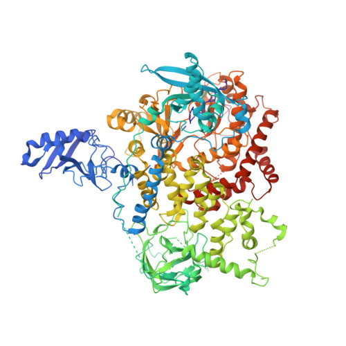
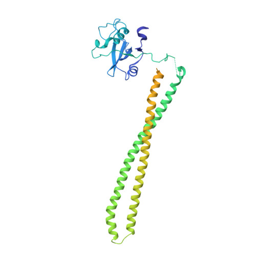
A frequent kinase domain mutation that changes the interaction between PI3K{alpha} and the membrane.

Mandelker, D.Gabelli, S.B.Schmidt-Kittler, O.Zhu, J.Cheong, I.Huang, C.H.Kinzler, K.W.Vogelstein, B.Amzel, L.M.

(2009) Proc Natl Acad Sci U S A 106: 16996-17001

  • DOI: https://doi.org/10.1073/pnas.0908444106
  • Primary Citation of Related Structures:  
    3HHM, 3HIZ

  • PubMed Abstract: 

    Mutations in oncogenes often promote tumorigenesis by changing the conformation of the encoded proteins, thereby altering enzymatic activity. The PIK3CA oncogene, which encodes p110alpha, the catalytic subunit of phosphatidylinositol 3-kinase alpha (PI3Kalpha), is one of the two most frequently mutated oncogenes in human cancers. We report the structure of the most common mutant of p110alpha in complex with two interacting domains of its regulatory partner (p85alpha), both free and bound to an inhibitor (wortmannin). The N-terminal SH2 (nSH2) domain of p85alpha is shown to form a scaffold for the entire enzyme complex, strategically positioned to communicate extrinsic signals from phosphopeptides to three distinct regions of p110alpha. Moreover, we found that Arg-1047 points toward the cell membrane, perpendicular to the orientation of His-1047 in the WT enzyme. Surprisingly, two loops of the kinase domain that contact the cell membrane shift conformation in the oncogenic mutant. Biochemical assays revealed that the enzymatic activity of the p110alpha His1047Arg mutant is differentially regulated by lipid membrane composition. These structural and biochemical data suggest a previously undescribed mechanism for mutational activation of a kinase that involves perturbation of its interaction with the cellular membrane.


  • Organizational Affiliation

    The Ludwig Center for Cancer Genetics and Therapeutics and Howard Hughes Medical Institute, Johns Hopkins Kimmel Cancer Center, Baltimore, MD 21231, USA.


Macromolecules
Find similar proteins by:  (by identity cutoff)  |  3D Structure
Entity ID: 1
MoleculeChains Sequence LengthOrganismDetailsImage
Phosphatidylinositol-4,5-bisphosphate 3-kinase catalytic subunit alpha isoform1,091Homo sapiensMutation(s): 1 
Gene Names: PIK3CA
EC: 2.7.1.153
UniProt & NIH Common Fund Data Resources
Find proteins for P42336 (Homo sapiens)
Explore P42336 
Go to UniProtKB:  P42336
PHAROS:  P42336
GTEx:  ENSG00000121879 
Entity Groups  
Sequence Clusters30% Identity50% Identity70% Identity90% Identity95% Identity100% Identity
UniProt GroupP42336
Sequence Annotations
Expand
  • Reference Sequence
Find similar proteins by:  (by identity cutoff)  |  3D Structure
Entity ID: 2
MoleculeChains Sequence LengthOrganismDetailsImage
niSH2 p85alpha373Homo sapiensMutation(s): 1 
Gene Names: PIK3R1
UniProt & NIH Common Fund Data Resources
Find proteins for P27986 (Homo sapiens)
Explore P27986 
Go to UniProtKB:  P27986
PHAROS:  P27986
GTEx:  ENSG00000145675 
Entity Groups  
Sequence Clusters30% Identity50% Identity70% Identity90% Identity95% Identity100% Identity
UniProt GroupP27986
Sequence Annotations
Expand
  • Reference Sequence
Small Molecules
Ligands 1 Unique
IDChains Name / Formula / InChI Key2D Diagram3D Interactions
KWT
Query on KWT

Download Ideal Coordinates CCD File 
C [auth A](1S,6BR,9AS,11R,11BR)-9A,11B-DIMETHYL-1-[(METHYLOXY)METHYL]-3,6,9-TRIOXO-1,6,6B,7,8,9,9A,10,11,11B-DECAHYDRO-3H-FURO[4, 3,2-DE]INDENO[4,5-H][2]BENZOPYRAN-11-YL ACETATE
C23 H24 O8
QDLHCMPXEPAAMD-QAIWCSMKSA-N
Binding Affinity Annotations 
IDSourceBinding Affinity
KWT BindingDB:  3HHM Ki: 120 (nM) from 1 assay(s)
Kd: 5.4 (nM) from 1 assay(s)
IC50: min: 1, max: 1610 (nM) from 11 assay(s)
PDBBind:  3HHM IC50: 11.9 (nM) from 1 assay(s)
Experimental Data & Validation

Experimental Data

  • Method: X-RAY DIFFRACTION
  • Resolution: 2.80 Å
  • R-Value Free: 0.307 
  • R-Value Work: 0.223 
  • R-Value Observed: 0.239 
  • Space Group: P 21 21 21
Unit Cell:
Length ( Å )Angle ( ˚ )
a = 115.312α = 90
b = 121.453β = 90
c = 152.494γ = 90
Software Package:
Software NamePurpose
DENZOdata reduction
SCALEPACKdata scaling
REFMACrefinement
PDB_EXTRACTdata extraction
MAR345data collection
REFMACphasing

Structure Validation

View Full Validation Report


Entry History 

Deposition Data

Revision History  (Full details and data files)

  • Version 1.0: 2009-09-29
    Type: Initial release
  • Version 1.1: 2011-07-13
    Changes: Version format compliance
  • Version 1.2: 2017-11-01
    Changes: Refinement description
  • Version 1.3: 2018-09-26
    Changes: Data collection, Source and taxonomy
  • Version 1.4: 2021-10-13
    Changes: Database references, Derived calculations, Structure summary
  • Version 1.5: 2023-09-06
    Changes: Data collection, Refinement description