3GEF

Crystal structure of the R482W mutant of lamin A/C


Experimental Data Snapshot

  • Method: X-RAY DIFFRACTION
  • Resolution: 1.50 Å
  • R-Value Free: 0.253 
  • R-Value Work: 0.214 

wwPDB Validation   3D Report Full Report


This is version 1.4 of the entry. See complete history


Literature

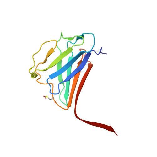
Structure of the lamin A/C R482W mutant responsible for dominant familial partial lipodystrophy (FPLD).

Magracheva, E.Kozlov, S.Stewart, C.L.Wlodawer, A.Zdanov, A.

(2009) Acta Crystallogr Sect F Struct Biol Cryst Commun 65: 665-670

  • DOI: https://doi.org/10.1107/S1744309109020302
  • Primary Citation of Related Structures:  
    3GEF

  • PubMed Abstract: 

    Proteins of the A-type lamin family, which consists of two members, lamin A and lamin C, are the major components of a thin proteinaceous filamentous meshwork, the lamina, that underlies the inner nuclear membrane. A-type lamins have recently become the focus of extensive functional studies as a consequence of the linking of at least eight congenital diseases to mutations in the lamin A/C gene (LMNA). This spectrum of pathologies, which mostly manifest themselves as dominant traits, includes muscle dystrophies, dilated cardiomyopathies, the premature aging syndrome Hutchinson-Guilford progeria and familial partial lipodystrophy (FPLD). The crystal structure of the lamin A/C mutant R482W, a variant that causes FPLD, has been determined at 1.5 A resolution. A completely novel aggregation state of the C-terminal globular domain and the position of the mutated amino-acid residue suggest means by which the mutation may affect lamin A/C-protein and protein-DNA interactions.


  • Organizational Affiliation

    Basic Research Program SAIC-Frederick, NCI-Frederick, Frederick, MD 21702, USA.


Macromolecules
Find similar proteins by:  (by identity cutoff)  |  3D Structure
Entity ID: 1
MoleculeChains Sequence LengthOrganismDetailsImage
Lamin-A/C
A, B, C, D
118Homo sapiensMutation(s): 1 
Gene Names: Lamin A/C 435-552LMN1LMNA
UniProt & NIH Common Fund Data Resources
Find proteins for P02545 (Homo sapiens)
Explore P02545 
Go to UniProtKB:  P02545
PHAROS:  P02545
GTEx:  ENSG00000160789 
Entity Groups  
Sequence Clusters30% Identity50% Identity70% Identity90% Identity95% Identity100% Identity
UniProt GroupP02545
Sequence Annotations
Expand
  • Reference Sequence
Small Molecules
Modified Residues  1 Unique
IDChains TypeFormula2D DiagramParent
CSD
Query on CSD
A, B, C, D
L-PEPTIDE LINKINGC3 H7 N O4 SCYS
Experimental Data & Validation

Experimental Data

  • Method: X-RAY DIFFRACTION
  • Resolution: 1.50 Å
  • R-Value Free: 0.253 
  • R-Value Work: 0.214 
  • Space Group: P 21 21 21
Unit Cell:
Length ( Å )Angle ( ˚ )
a = 62.195α = 90
b = 84.035β = 90
c = 98.75γ = 90
Software Package:
Software NamePurpose
HKL-2000data collection
PHASERphasing
CNSrefinement
DENZOdata reduction
SCALEPACKdata scaling

Structure Validation

View Full Validation Report


Entry History 

Deposition Data

Revision History  (Full details and data files)

  • Version 1.0: 2009-08-04
    Type: Initial release
  • Version 1.1: 2011-07-13
    Changes: Version format compliance
  • Version 1.2: 2014-11-19
    Changes: Non-polymer description
  • Version 1.3: 2021-10-20
    Changes: Database references, Derived calculations
  • Version 1.4: 2023-09-06
    Changes: Data collection, Refinement description