3FBX

Crystal structure of the lysosomal 66.3 kDa protein from mouse solved by S-SAD


Experimental Data Snapshot

  • Method: X-RAY DIFFRACTION
  • Resolution: 2.40 Å
  • R-Value Free: 0.198 
  • R-Value Work: 0.156 
  • R-Value Observed: 0.158 

wwPDB Validation   3D Report Full Report


Ligand Structure Quality Assessment 


This is version 2.1 of the entry. See complete history


Literature

De novo sulfur SAD phasing of the lysosomal 66.3 kDa protein from mouse

Lakomek, K.Dickmanns, A.Mueller, U.Kollmann, K.Deuschl, F.Berndt, A.Lubke, T.Ficner, R.

(2009) Acta Crystallogr D Biol Crystallogr 65: 220-228

  • DOI: https://doi.org/10.1107/S0907444908041814
  • Primary Citation of Related Structures:  
    3FBX

  • PubMed Abstract: 

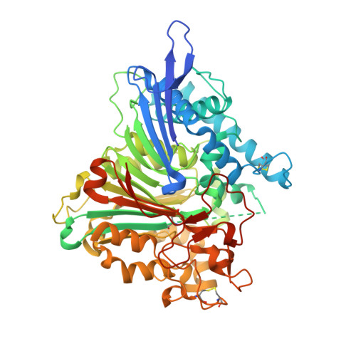
    The 66.3 kDa protein from mouse is a soluble protein of the lysosomal matrix. It is synthesized as a glycosylated 75 kDa preproprotein which is further processed into 28 and 40 kDa fragments. Despite bioinformatics approaches and molecular characterization of the 66.3 kDa protein, the mode of its maturation as well as its physiological function remained unknown. Therefore, it was decided to tackle this question by means of X-ray crystallography. After expression in a human fibrosarcoma cell line, the C-terminally His-tagged single-chain 66.3 kDa variant and the double-chain form consisting of a 28 kDa fragment and a 40 kDa fragment were purified to homogeneity but could not be separated during the purification procedure. This mixture was therefore used for crystallization. Single crystals were obtained and the structure of the 66.3 kDa protein was solved by means of sulfur SAD phasing using data collected at a wavelength of 1.9 A on the BESSY beamline BL14.2 of Freie Universität Berlin. Based on the anomalous signal, a 22-atom substructure comprising 21 intrinsic S atoms and one Xe atom with very low occupancy was found and refined at a resolution of 2.4 A using the programs SHELXC/D and SHARP. Density modification using SOLOMON and DM resulted in a high-quality electron-density map, enabling automatic model building with ARP/wARP. The initial model contained 85% of the amino-acid residues expected to be present in the asymmetric unit of the crystal. Subsequently, the model was completed and refined to an R(free) factor of 19.8%. The contribution of the single Xe atom to the anomalous signal was analyzed in comparison to that of the S atoms and was found to be negligible. This work should encourage the use of the weak anomalous scattering of intrinsic S atoms in SAD phasing, especially for proteins, which require both expensive and time-consuming expression and purification procedures, preventing extensive screening of heavy-atom crystal soaks.


  • Organizational Affiliation

    Department of Molecular Structural Biology, Institute of Microbiology and Genetics, Georg-August University Göttingen, Göttingen, Germany.


Macromolecules
Find similar proteins by:  (by identity cutoff)  |  3D Structure
Entity ID: 1
MoleculeChains Sequence LengthOrganismDetailsImage
Putative phospholipase B-like 2559Mus musculusMutation(s): 0 
Gene Names: AAG44101
EC: 3.1.1
UniProt & NIH Common Fund Data Resources
Find proteins for Q3TCN2 (Mus musculus)
Explore Q3TCN2 
Go to UniProtKB:  Q3TCN2
IMPC:  MGI:1919022
Entity Groups  
Sequence Clusters30% Identity50% Identity70% Identity90% Identity95% Identity100% Identity
UniProt GroupQ3TCN2
Sequence Annotations
Expand
  • Reference Sequence
Oligosaccharides

Help

Entity ID: 2
MoleculeChains Length2D Diagram Glycosylation3D Interactions
2-acetamido-2-deoxy-beta-D-glucopyranose-(1-4)-2-acetamido-2-deoxy-beta-D-glucopyranose
B
2N-Glycosylation
Glycosylation Resources
GlyTouCan:  G42666HT
GlyCosmos:  G42666HT
GlyGen:  G42666HT
Small Molecules
Ligands 7 Unique
IDChains Name / Formula / InChI Key2D Diagram3D Interactions
NAG
Query on NAG

Download Ideal Coordinates CCD File 
C [auth A],
D [auth A],
E [auth A]
2-acetamido-2-deoxy-beta-D-glucopyranose
C8 H15 N O6
OVRNDRQMDRJTHS-FMDGEEDCSA-N
PG4
Query on PG4

Download Ideal Coordinates CCD File 
F [auth A],
G [auth A]
TETRAETHYLENE GLYCOL
C8 H18 O5
UWHCKJMYHZGTIT-UHFFFAOYSA-N
PGE
Query on PGE

Download Ideal Coordinates CCD File 
M [auth A]TRIETHYLENE GLYCOL
C6 H14 O4
ZIBGPFATKBEMQZ-UHFFFAOYSA-N
XE
Query on XE

Download Ideal Coordinates CCD File 
H [auth A]XENON
Xe
FHNFHKCVQCLJFQ-UHFFFAOYSA-N
GOL
Query on GOL

Download Ideal Coordinates CCD File 
I [auth A],
K [auth A],
L [auth A]
GLYCEROL
C3 H8 O3
PEDCQBHIVMGVHV-UHFFFAOYSA-N
ACT
Query on ACT

Download Ideal Coordinates CCD File 
J [auth A]ACETATE ION
C2 H3 O2
QTBSBXVTEAMEQO-UHFFFAOYSA-M
NA
Query on NA

Download Ideal Coordinates CCD File 
N [auth A]SODIUM ION
Na
FKNQFGJONOIPTF-UHFFFAOYSA-N
Modified Residues  1 Unique
IDChains TypeFormula2D DiagramParent
OCS
Query on OCS
A
L-PEPTIDE LINKINGC3 H7 N O5 SCYS
Experimental Data & Validation

Experimental Data

  • Method: X-RAY DIFFRACTION
  • Resolution: 2.40 Å
  • R-Value Free: 0.198 
  • R-Value Work: 0.156 
  • R-Value Observed: 0.158 
  • Space Group: C 1 2 1
Unit Cell:
Length ( Å )Angle ( ˚ )
a = 148.804α = 90
b = 89.672β = 98.67
c = 64.954γ = 90
Software Package:
Software NamePurpose
DENZOdata reduction
SCALEPACKdata scaling
SHARPphasing
REFMACrefinement
PDB_EXTRACTdata extraction
MAR345dtbdata collection
HKL-2000data reduction
HKL-2000data scaling

Structure Validation

View Full Validation Report


Ligand Structure Quality Assessment 


Entry History 

Deposition Data

Revision History  (Full details and data files)

  • Version 1.0: 2009-03-03
    Type: Initial release
  • Version 1.1: 2011-07-13
    Changes: Non-polymer description, Version format compliance
  • Version 1.2: 2017-11-01
    Changes: Refinement description
  • Version 2.0: 2020-07-29
    Type: Remediation
    Reason: Carbohydrate remediation
    Changes: Atomic model, Data collection, Database references, Derived calculations, Structure summary
  • Version 2.1: 2023-12-27
    Changes: Data collection, Database references, Structure summary