3DIW

c-terminal beta-catenin bound TIP-1 structure


Experimental Data Snapshot

  • Method: X-RAY DIFFRACTION
  • Resolution: 2.10 Å
  • R-Value Free: 0.265 
  • R-Value Work: 0.221 
  • R-Value Observed: 0.221 

wwPDB Validation   3D Report Full Report


This is version 1.3 of the entry. See complete history


Literature

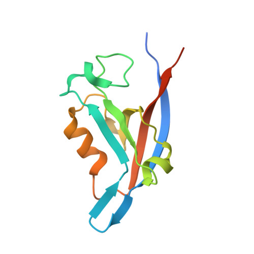
Structural Basis of beta-Catenin Recognition by Tax-interacting Protein-1

Zhang, J.Yan, X.Shi, C.Yang, X.Guo, Y.Tian, C.Long, J.Shen, Y.

(2008) J Mol Biol 384: 255-263

  • DOI: https://doi.org/10.1016/j.jmb.2008.09.034
  • Primary Citation of Related Structures:  
    3DIW, 3DJ1, 3DJ3

  • PubMed Abstract: 

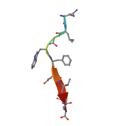
    Tax-interacting protein-1 (TIP-1) is an unusual signaling protein, containing a single PDZ domain. TIP-1 is able to bind beta-catenin with high affinity and thus inhibit its transcriptional activity. The high-resolution crystal structure of TIP-1 in complex with the C-terminal peptide of beta-catenin provides molecular details for the recognition of beta-catenin by TIP-1. Moreover, structural comparison of peptide-free and peptide-bound TIP-1 reveals that significant conformational changes are required in the betaB-betaC loop region of TIP-1 to avoid clashes with the incoming C-terminal beta-catenin peptide. Such conformational changes have not been observed in other structures of PDZ domains. In addition to the canonical peptide-binding pocket of the PDZ domain, TIP-1 can form a binding cavity to anchor more amino acids through a conserved hydrophobic residue pair (Trp776 of beta-catenin and Pro45 of TIP-1). Structural and biochemical data indicate that the canonical binding pocket together with the hydrophobic residue pair are presumably the major cause of the significantly higher affinity of the beta-catenin C-terminal to TIP-1 than to other PDZ domains, providing a unique binding specificity. Our results reveal the molecular mechanism of TIP-1 as an antagonist in PDZ domain signaling.


  • Organizational Affiliation

    Tianjin Key Laboratory of Protein Science, College of Life Science, NanKai University, Tianjin 300071, China.


Macromolecules
Find similar proteins by:  (by identity cutoff)  |  3D Structure
Entity ID: 1
MoleculeChains Sequence LengthOrganismDetailsImage
Tax1-binding protein 3
A, B
124Mus musculusMutation(s): 0 
UniProt & NIH Common Fund Data Resources
Find proteins for Q9DBG9 (Mus musculus)
Explore Q9DBG9 
Go to UniProtKB:  Q9DBG9
IMPC:  MGI:1923531
Entity Groups  
Sequence Clusters30% Identity50% Identity70% Identity90% Identity95% Identity100% Identity
UniProt GroupQ9DBG9
Sequence Annotations
Expand
  • Reference Sequence

Find similar proteins by:  Sequence   |   3D Structure  

Entity ID: 2
MoleculeChains Sequence LengthOrganismDetailsImage
decameric peptide form Catenin beta-1
C, D
10N/AMutation(s): 0 
UniProt & NIH Common Fund Data Resources
Find proteins for P35222 (Homo sapiens)
Explore P35222 
Go to UniProtKB:  P35222
PHAROS:  P35222
GTEx:  ENSG00000168036 
Entity Groups  
Sequence Clusters30% Identity50% Identity70% Identity90% Identity95% Identity100% Identity
UniProt GroupP35222
Sequence Annotations
Expand
  • Reference Sequence
Experimental Data & Validation

Experimental Data

  • Method: X-RAY DIFFRACTION
  • Resolution: 2.10 Å
  • R-Value Free: 0.265 
  • R-Value Work: 0.221 
  • R-Value Observed: 0.221 
  • Space Group: P 21 21 21
Unit Cell:
Length ( Å )Angle ( ˚ )
a = 27.098α = 90
b = 93.571β = 90
c = 107.724γ = 90
Software Package:
Software NamePurpose
CNSrefinement
HKL-2000data collection
HKL-2000data reduction
SCALEPACKdata scaling
PHASESphasing

Structure Validation

View Full Validation Report


Entry History 

Deposition Data

  • Released Date: 2008-10-21 
  • Deposition Author(s): Shen, Y.

Revision History  (Full details and data files)

  • Version 1.0: 2008-10-21
    Type: Initial release
  • Version 1.1: 2011-07-13
    Changes: Version format compliance
  • Version 1.2: 2014-02-05
    Changes: Derived calculations
  • Version 1.3: 2023-11-01
    Changes: Data collection, Database references, Refinement description