3DAB

Structure of the human Mdmx protein bound to the p53 tumor suppressor transactivation domain


Experimental Data Snapshot

  • Method: X-RAY DIFFRACTION
  • Resolution: 1.90 Å
  • R-Value Free: 0.257 
  • R-Value Work: 0.197 
  • R-Value Observed: 0.200 

wwPDB Validation   3D Report Full Report


This is version 1.4 of the entry. See complete history


Literature

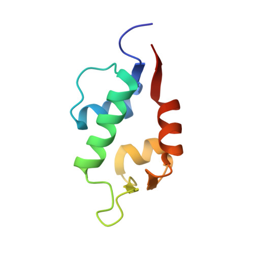
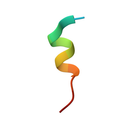
Structure of the human Mdmx protein bound to the p53 tumor suppressor transactivation domain.

Popowicz, G.M.Czarna, A.Holak, T.A.

(2008) Cell Cycle 7: 2441-2443

  • DOI: https://doi.org/10.4161/cc.6365
  • Primary Citation of Related Structures:  
    3DAB, 3DAC

  • PubMed Abstract: 

    The Mdmx oncoprotein has only recently emerged as a critical-independent to Mdm2-regulator of p53 activation. We have determined the crystal structure of the N-terminal domain of human Mdmx bound to a 15-residue transactivation domain peptide of human p53. The structure shows why antagonists of the Mdm2 binding to p53 are ineffective in the Mdmx-p53 interaction.


Macromolecules
Find similar proteins by:  (by identity cutoff)  |  3D Structure
Entity ID: 1
MoleculeChains Sequence LengthOrganismDetailsImage
Mdm4 protein
A, C, E, G
90Homo sapiensMutation(s): 0 
Gene Names: MDM4MDMX
UniProt & NIH Common Fund Data Resources
Find proteins for O15151 (Homo sapiens)
Explore O15151 
Go to UniProtKB:  O15151
PHAROS:  O15151
GTEx:  ENSG00000198625 
Entity Groups  
Sequence Clusters30% Identity50% Identity70% Identity90% Identity95% Identity100% Identity
UniProt GroupO15151
Sequence Annotations
Expand
  • Reference Sequence

Find similar proteins by:  Sequence   |   3D Structure  

Entity ID: 2
MoleculeChains Sequence LengthOrganismDetailsImage
Cellular tumor antigen p53
B, D, F, H
15N/AMutation(s): 0 
UniProt & NIH Common Fund Data Resources
Find proteins for P04637 (Homo sapiens)
Explore P04637 
Go to UniProtKB:  P04637
PHAROS:  P04637
GTEx:  ENSG00000141510 
Entity Groups  
Sequence Clusters30% Identity50% Identity70% Identity90% Identity95% Identity100% Identity
UniProt GroupP04637
Sequence Annotations
Expand
  • Reference Sequence
Experimental Data & Validation

Experimental Data

  • Method: X-RAY DIFFRACTION
  • Resolution: 1.90 Å
  • R-Value Free: 0.257 
  • R-Value Work: 0.197 
  • R-Value Observed: 0.200 
  • Space Group: P 1 21 1
Unit Cell:
Length ( Å )Angle ( ˚ )
a = 36.73α = 90
b = 58.87β = 92
c = 96.11γ = 90
Software Package:
Software NamePurpose
REFMACrefinement
MAR345data collection
XDSdata reduction
XSCALEdata scaling
MOLREPphasing

Structure Validation

View Full Validation Report


Entry History 

Deposition Data

Revision History  (Full details and data files)

  • Version 1.0: 2008-09-02
    Type: Initial release
  • Version 1.1: 2011-07-13
    Changes: Version format compliance
  • Version 1.2: 2017-10-25
    Changes: Refinement description
  • Version 1.3: 2019-11-06
    Changes: Data collection, Database references
  • Version 1.4: 2023-11-01
    Changes: Data collection, Database references, Refinement description