3CS6

Structure-based design of a superagonist ligand for the vitamin D nuclear receptor


Experimental Data Snapshot

  • Method: X-RAY DIFFRACTION
  • Resolution: 1.80 Å
  • R-Value Free: 0.206 
  • R-Value Work: 0.192 
  • R-Value Observed: 0.192 

wwPDB Validation   3D Report Full Report


Ligand Structure Quality Assessment 


This is version 1.2 of the entry. See complete history


Literature

Structure-based design of a superagonist ligand for the vitamin d nuclear receptor.

Hourai, S.Rodrigues, L.C.Antony, P.Reina-San-Martin, B.Ciesielski, F.Magnier, B.C.Schoonjans, K.Mourino, A.Rochel, N.Moras, D.

(2008) Chem Biol 15: 383-392

  • DOI: https://doi.org/10.1016/j.chembiol.2008.03.016
  • Primary Citation of Related Structures:  
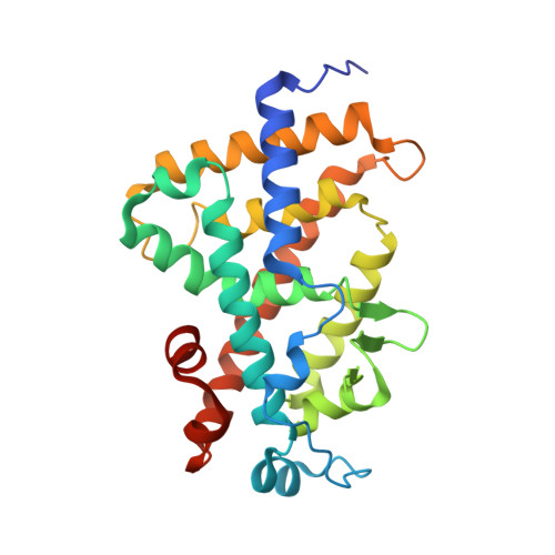
    3CS4, 3CS6

  • PubMed Abstract: 

    Vitamin D nuclear receptor (VDR), a ligand-dependent transcriptional regulator, is an important target for multiple clinical applications, such as osteoporosis and cancer. Since exacerbated increase of calcium serum level is currently associated with VDR ligands action, superagonists with low calcium serum levels have been developed. Based on the crystal structures of human VDR (hVDR) bound to 1alpha,25-dihydroxyvitamin D(3) and superagonists-notably, KH1060-we designed a superagonist ligand. In order to optimize the aliphatic side chain conformation with a subsequent entropy benefit, we incorporated an oxolane ring and generated two stereo diasteromers, AMCR277A and AMCR277B. Only AMCR277A exhibits superagonist activity in vitro, but is as calcemic in vivo as the natural ligand. The crystal structures of the complexes between the ligand binding domain of hVDR and these ligands provide a rational approach to the design of more potent superagonist ligands for potential clinical application.


  • Organizational Affiliation

    Institut de Génétique et de Biologie Moléculaire et Cellulaire, Département de Biologie et de Génomique Structurales, Université Louis Pasteur, Strasbourg F-67000, France.


Macromolecules
Find similar proteins by:  (by identity cutoff)  |  3D Structure
Entity ID: 1
MoleculeChains Sequence LengthOrganismDetailsImage
Vitamin D3 receptor263Homo sapiensMutation(s): 0 
Gene Names: VDRNR1I1
UniProt & NIH Common Fund Data Resources
Find proteins for P11473 (Homo sapiens)
Explore P11473 
Go to UniProtKB:  P11473
PHAROS:  P11473
GTEx:  ENSG00000111424 
Entity Groups  
Sequence Clusters30% Identity50% Identity70% Identity90% Identity95% Identity100% Identity
UniProt GroupP11473
Sequence Annotations
Expand
  • Reference Sequence
Small Molecules
Ligands 1 Unique
IDChains Name / Formula / InChI Key2D Diagram3D Interactions
0CO
Query on 0CO

Download Ideal Coordinates CCD File 
B [auth A](1S,3R,5Z,7E,14beta,17alpha,23R)-23-(2-hydroxy-2-methylpropyl)-20,24-epoxy-9,10-secochola-5,7,10-triene-1,3-diol
C28 H44 O4
QFEREDUWILIRPI-FOPXFMLJSA-N
Experimental Data & Validation

Experimental Data

  • Method: X-RAY DIFFRACTION
  • Resolution: 1.80 Å
  • R-Value Free: 0.206 
  • R-Value Work: 0.192 
  • R-Value Observed: 0.192 
  • Space Group: P 21 21 21
Unit Cell:
Length ( Å )Angle ( ˚ )
a = 45.055α = 90
b = 51.458β = 90
c = 132.132γ = 90
Software Package:
Software NamePurpose
ADSCdata collection
CNSrefinement
HKL-2000data reduction
HKL-2000data scaling
CNSphasing

Structure Validation

View Full Validation Report


Ligand Structure Quality Assessment 


Entry History 

Deposition Data

Revision History  (Full details and data files)

  • Version 1.0: 2008-05-27
    Type: Initial release
  • Version 1.1: 2011-07-13
    Changes: Version format compliance
  • Version 1.2: 2024-02-21
    Changes: Data collection, Database references, Derived calculations