3B9K

Crystal structure of CD8alpha-beta in complex with YTS 156.7 FAB


Experimental Data Snapshot

  • Method: X-RAY DIFFRACTION
  • Resolution: 2.70 Å
  • R-Value Free: 0.284 
  • R-Value Work: 0.235 
  • R-Value Observed: 0.238 

wwPDB Validation   3D Report Full Report


Ligand Structure Quality Assessment 


This is version 1.5 of the entry. See complete history


Literature

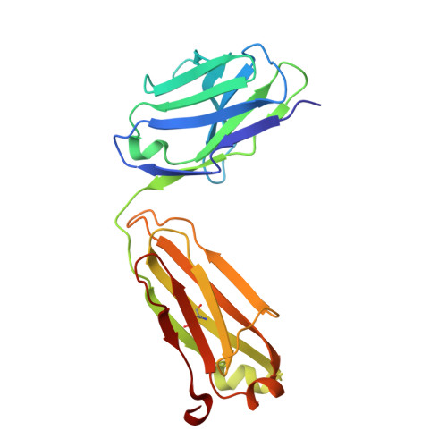
The Crystal Structure of CD8 in Complex with YTS156.7.7 Fab and Interaction with Other CD8 Antibodies Define the Binding Mode of CD8 alphabeta to MHC Class I

Shore, D.A.Issafras, H.Landais, E.Teyton, L.Wilson, I.A.

(2008) J Mol Biol 384: 1190-1202

  • DOI: https://doi.org/10.1016/j.jmb.2008.09.069
  • Primary Citation of Related Structures:  
    3B9K

  • PubMed Abstract: 

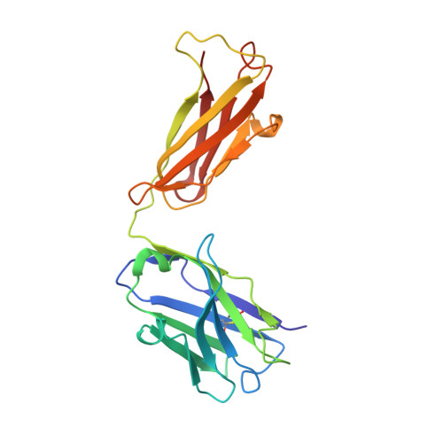
    The CD8alphabeta heterodimer interacts with class I pMHC on antigen-presenting cells as a co-receptor for TCR-mediated activation of cytotoxic T cells. To characterize this immunologically important interaction, we used monoclonal antibodies (mAbs) specific to either CD8alpha or CD8beta to probe the mechanism of CD8alphabeta binding to pMHCI. The YTS156.7 mAb inhibits this interaction and blocks T cell activation. To elucidate the molecular basis for this inhibition, the crystal structure of the CD8alphabeta immunoglobulin-like ectodomains were determined in complex with mAb YTS156.7 Fab at 2.7 A resolution. The YTS156.7 epitope on CD8beta was identified and implies that residues in the CDR1 and CDR2-equivalent loops of CD8beta are occluded upon binding to class I pMHC. To further characterize the pMHCI/CD8alphabeta interaction, binding of class I tetramers to CD8alphabeta on the surface of T cells was assessed in the presence of anti-CD8 mAbs. In contrast to YTS156.7, mAb YTS105.18, which is specific for CD8alpha, does not inhibit binding of CD8alphabeta to class I tetramers, indicating the YTS105.18 epitope is not occluded in the pMHCI/CD8alphabeta complex. Together, these data indicate a model for the pMHCI/CD8alphabeta interaction similar to that observed for CD8alphaalpha in the CD8alphaalpha/pMHCI complex, but in which CD8alpha occupies the lower orientation (membrane proximal to the antigen presenting cell), and CD8beta occupies the upper position (membrane distal). The implication of this molecular assembly for the function of CD8alphabeta in T cell activation is discussed.


  • Organizational Affiliation

    Department of Molecular Biology, The Scripps Research Institute, La Jolla, CA 92037, USA.


Macromolecules
Find similar proteins by:  (by identity cutoff)  |  3D Structure
Entity ID: 1
MoleculeChains Sequence LengthOrganismDetailsImage
Fab Light ChainA [auth L],
E [auth C]
213Rattus rattusMutation(s): 0 
Entity Groups  
Sequence Clusters30% Identity50% Identity70% Identity90% Identity95% Identity100% Identity
Sequence Annotations
Expand
  • Reference Sequence
Find similar proteins by:  (by identity cutoff)  |  3D Structure
Entity ID: 2
MoleculeChains Sequence LengthOrganismDetailsImage
Fab Heavy chainB [auth H],
F [auth D]
214Rattus rattusMutation(s): 0 
Entity Groups  
Sequence Clusters30% Identity50% Identity70% Identity90% Identity95% Identity100% Identity
Sequence Annotations
Expand
  • Reference Sequence
Find similar proteins by:  (by identity cutoff)  |  3D Structure
Entity ID: 3
MoleculeChains Sequence LengthOrganismDetailsImage
T-cell surface glycoprotein CD8 alpha chainC [auth A],
G [auth E]
131Mus musculusMutation(s): 0 
Gene Names: Cd8aLyt-2Lyt2
UniProt
Find proteins for P01731 (Mus musculus)
Explore P01731 
Go to UniProtKB:  P01731
Entity Groups  
Sequence Clusters30% Identity50% Identity70% Identity90% Identity95% Identity100% Identity
UniProt GroupP01731
Sequence Annotations
Expand
  • Reference Sequence
Find similar proteins by:  (by identity cutoff)  |  3D Structure
Entity ID: 4
MoleculeChains Sequence LengthOrganismDetailsImage
T-cell surface glycoprotein CD8 beta chainD [auth B],
H [auth F]
125Mus musculusMutation(s): 0 
Gene Names: Cd8bCd8b1Ly-3Lyt-3Lyt3
UniProt
Find proteins for P10300 (Mus musculus)
Explore P10300 
Go to UniProtKB:  P10300
Entity Groups  
Sequence Clusters30% Identity50% Identity70% Identity90% Identity95% Identity100% Identity
UniProt GroupP10300
Sequence Annotations
Expand
  • Reference Sequence
Small Molecules
Ligands 1 Unique
IDChains Name / Formula / InChI Key2D Diagram3D Interactions
NAG
Query on NAG

Download Ideal Coordinates CCD File 
I [auth A],
J [auth E]
2-acetamido-2-deoxy-beta-D-glucopyranose
C8 H15 N O6
OVRNDRQMDRJTHS-FMDGEEDCSA-N
Experimental Data & Validation

Experimental Data

  • Method: X-RAY DIFFRACTION
  • Resolution: 2.70 Å
  • R-Value Free: 0.284 
  • R-Value Work: 0.235 
  • R-Value Observed: 0.238 
  • Space Group: P 21 21 21
Unit Cell:
Length ( Å )Angle ( ˚ )
a = 91.779α = 90
b = 92.747β = 90
c = 190.31γ = 90
Software Package:
Software NamePurpose
DENZOdata reduction
SCALEPACKdata scaling
PHASERphasing
REFMACrefinement
PDB_EXTRACTdata extraction
HKL-2000data collection
HKL-2000data reduction

Structure Validation

View Full Validation Report


Ligand Structure Quality Assessment 


Entry History 

Deposition Data

Revision History  (Full details and data files)

  • Version 1.0: 2008-11-11
    Type: Initial release
  • Version 1.1: 2011-07-13
    Changes: Non-polymer description, Version format compliance
  • Version 1.2: 2013-09-25
    Changes: Derived calculations
  • Version 1.3: 2016-06-22
    Changes: Structure summary
  • Version 1.4: 2017-10-25
    Changes: Refinement description
  • Version 1.5: 2020-07-29
    Type: Remediation
    Reason: Carbohydrate remediation
    Changes: Data collection, Database references, Derived calculations, Structure summary