3B2V

Crystal structure of the extracellular region of the epidermal growth factor receptor in complex with the Fab fragment of IMC-11F8


Experimental Data Snapshot

  • Method: X-RAY DIFFRACTION
  • Resolution: 3.30 Å
  • R-Value Free: 0.367 
  • R-Value Work: 0.282 
  • R-Value Observed: 0.286 

wwPDB Validation   3D Report Full Report


Ligand Structure Quality Assessment 


This is version 2.1 of the entry. See complete history


Literature

Structural basis for EGF receptor inhibition by the therapeutic antibody IMC-11F8.

Li, S.Kussie, P.Ferguson, K.M.

(2008) Structure 16: 216-227

  • DOI: https://doi.org/10.1016/j.str.2007.11.009
  • Primary Citation of Related Structures:  
    3B2U, 3B2V

  • PubMed Abstract: 

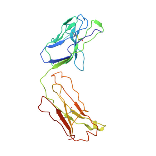
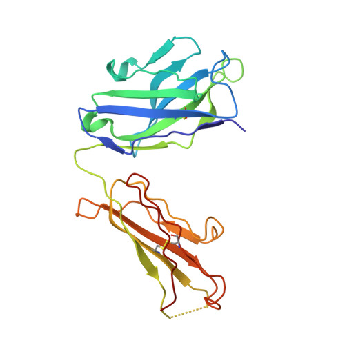
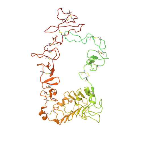
    Therapeutic anticancer strategies that target and inactivate the epidermal growth factor receptor (EGFR) are under intense study in the clinic. Here we describe the mechanism of EGFR inhibition by an antibody drug IMC-11F8. IMC-11F8 is a fully human antibody that has similar antitumor potency as the chimeric cetuximab/Erbitux and might represent a safer therapeutic alternative. We report the X-ray crystal structure of the Fab fragment of IMC-11F8 (Fab11F8) in complex with the entire extracellular region and with isolated domain III of EGFR. We compare this to our previous study of the cetuximab/EGFR interaction. Fab11F8 interacts with a remarkably similar epitope, but through a completely different set of interactions. Both the similarities and differences in binding of these two antibodies have important implications for the development of inhibitors that could exploit this same mechanism of EGFR inhibition.


  • Organizational Affiliation

    Department of Physiology, University of Pennsylvania School of Medicine, Philadelphia, PA 19104, USA.


Macromolecules
Find similar proteins by:  (by identity cutoff)  |  3D Structure
Entity ID: 1
MoleculeChains Sequence LengthOrganismDetailsImage
IMC-11F8 FAB Light chainA [auth L]213Homo sapiensMutation(s): 0 
Gene Names: EGFR
Entity Groups  
Sequence Clusters30% Identity50% Identity70% Identity90% Identity95% Identity100% Identity
Sequence Annotations
Expand
  • Reference Sequence
Find similar proteins by:  (by identity cutoff)  |  3D Structure
Entity ID: 2
MoleculeChains Sequence LengthOrganismDetailsImage
IMC-11F8 FAB Heavy chainB [auth H]223Homo sapiensMutation(s): 0 
UniProt
Find proteins for Q6GMX6 (Homo sapiens)
Explore Q6GMX6 
Go to UniProtKB:  Q6GMX6
Entity Groups  
Sequence Clusters30% Identity50% Identity70% Identity90% Identity95% Identity100% Identity
UniProt GroupQ6GMX6
Sequence Annotations
Expand
  • Reference Sequence
Find similar proteins by:  (by identity cutoff)  |  3D Structure
Entity ID: 3
MoleculeChains Sequence LengthOrganismDetailsImage
Epidermal growth factor receptorC [auth A]624Homo sapiensMutation(s): 0 
Gene Names: EGFRERBB1
EC: 2.7.10.1
UniProt & NIH Common Fund Data Resources
Find proteins for P00533 (Homo sapiens)
Explore P00533 
Go to UniProtKB:  P00533
PHAROS:  P00533
GTEx:  ENSG00000146648 
Entity Groups  
Sequence Clusters30% Identity50% Identity70% Identity90% Identity95% Identity100% Identity
UniProt GroupP00533
Sequence Annotations
Expand
  • Reference Sequence
Oligosaccharides

Help

Entity ID: 4
MoleculeChains Length2D Diagram Glycosylation3D Interactions
beta-D-mannopyranose-(1-4)-2-acetamido-2-deoxy-beta-D-glucopyranose-(1-4)-2-acetamido-2-deoxy-beta-D-glucopyranoseD [auth B]3N-Glycosylation
Glycosylation Resources
GlyTouCan:  G15407YE
GlyCosmos:  G15407YE
GlyGen:  G15407YE
Entity ID: 5
MoleculeChains Length2D Diagram Glycosylation3D Interactions
2-acetamido-2-deoxy-beta-D-glucopyranose-(1-4)-2-acetamido-2-deoxy-beta-D-glucopyranoseE [auth C],
F [auth D]
2N-Glycosylation
Glycosylation Resources
GlyTouCan:  G42666HT
GlyCosmos:  G42666HT
GlyGen:  G42666HT
Small Molecules
Ligands 1 Unique
IDChains Name / Formula / InChI Key2D Diagram3D Interactions
NAG
Query on NAG

Download Ideal Coordinates CCD File 
G [auth A],
H [auth A]
2-acetamido-2-deoxy-beta-D-glucopyranose
C8 H15 N O6
OVRNDRQMDRJTHS-FMDGEEDCSA-N
Experimental Data & Validation

Experimental Data

  • Method: X-RAY DIFFRACTION
  • Resolution: 3.30 Å
  • R-Value Free: 0.367 
  • R-Value Work: 0.282 
  • R-Value Observed: 0.286 
  • Space Group: C 2 2 21
Unit Cell:
Length ( Å )Angle ( ˚ )
a = 68.743α = 90
b = 266.242β = 90
c = 156.381γ = 90
Software Package:
Software NamePurpose
DENZOdata reduction
SCALEPACKdata scaling
MOLREPphasing
DMphasing
REFMACrefinement
PDB_EXTRACTdata extraction
ADSCdata collection

Structure Validation

View Full Validation Report


Ligand Structure Quality Assessment 


Entry History 

Deposition Data

Revision History  (Full details and data files)

  • Version 1.0: 2008-02-19
    Type: Initial release
  • Version 1.1: 2011-07-13
    Changes: Non-polymer description, Version format compliance
  • Version 1.2: 2017-10-25
    Changes: Advisory, Refinement description
  • Version 2.0: 2020-07-29
    Type: Remediation
    Reason: Carbohydrate remediation
    Changes: Advisory, Atomic model, Data collection, Database references, Derived calculations, Non-polymer description, Structure summary
  • Version 2.1: 2023-08-30
    Changes: Data collection, Database references, Refinement description, Structure summary