3AL3

Crystal Structure of TopBP1 BRCT7/8-BACH1 peptide complex


Experimental Data Snapshot

  • Method: X-RAY DIFFRACTION
  • Resolution: 2.15 Å
  • R-Value Free: 0.236 
  • R-Value Work: 0.196 
  • R-Value Observed: 0.198 

wwPDB Validation   3D Report Full Report


This is version 1.4 of the entry. See complete history


Literature

Molecular basis of BACH1/FANCJ recognition by TopBP1 in DNA replication checkpoint control

Leung, C.C.Gong, Z.Chen, J.Glover, J.N.

(2011) J Biol Chem 286: 4292-4301

  • DOI: https://doi.org/10.1074/jbc.M110.189555
  • Primary Citation of Related Structures:  
    3AL2, 3AL3

  • PubMed Abstract: 

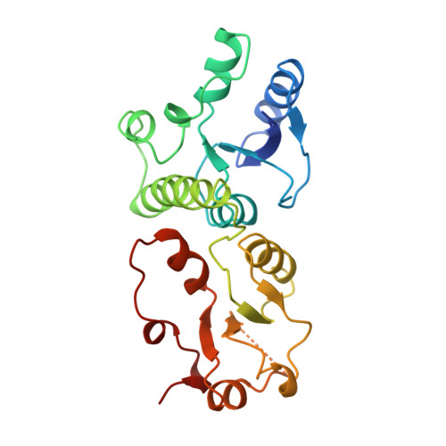
    The diverse roles of TopBP1 in DNA replication and checkpoint signaling are associated with the scaffolding ability of TopBP1 to initiate various protein-protein interactions. The recognition of the BACH1/FANCJ helicase by TopBP1 is critical for the activation of the DNA replication checkpoint at stalled replication forks and is facilitated by the C-terminal tandem BRCT7/8 domains of TopBP1 and a phosphorylated Thr(1133) binding motif in BACH1. Here we provide the structural basis for this interaction through analysis of the x-ray crystal structures of TopBP1 BRCT7/8 both free and in complex with a BACH1 phospho-peptide. In contrast to canonical BRCT-phospho-peptide recognition, TopBP1 BRCT7/8 undergoes a dramatic conformational change upon BACH1 binding such that the two BRCT repeats pivot about the central BRCT-BRCT interface to provide an extensive and deep peptide-binding cleft. Additionally, we provide the first structural mechanism for Thr(P) recognition among BRCT domains. Together with systematic mutagenesis studies, we highlight the role of key contacts in governing the unique specificity of the TopBP1-BACH1 interaction.


  • Organizational Affiliation

    Department of Biochemistry, School of Molecular and Systems Medicine, University of Alberta, Edmonton, Alberta T6G 2H7, Canada.


Macromolecules
Find similar proteins by:  (by identity cutoff)  |  3D Structure
Entity ID: 1
MoleculeChains Sequence LengthOrganismDetailsImage
DNA topoisomerase 2-binding protein 1235Homo sapiensMutation(s): 0 
Gene Names: KIAA0259TOPBP1
UniProt & NIH Common Fund Data Resources
Find proteins for Q92547 (Homo sapiens)
Explore Q92547 
Go to UniProtKB:  Q92547
PHAROS:  Q92547
GTEx:  ENSG00000163781 
Entity Groups  
Sequence Clusters30% Identity50% Identity70% Identity90% Identity95% Identity100% Identity
UniProt GroupQ92547
Sequence Annotations
Expand
  • Reference Sequence

Find similar proteins by:  Sequence   |   3D Structure  

Entity ID: 2
MoleculeChains Sequence LengthOrganismDetailsImage
Peptide of Fanconi anemia group J protein10N/AMutation(s): 0 
UniProt & NIH Common Fund Data Resources
Find proteins for Q9BX63 (Homo sapiens)
Explore Q9BX63 
Go to UniProtKB:  Q9BX63
PHAROS:  Q9BX63
GTEx:  ENSG00000136492 
Entity Groups  
Sequence Clusters30% Identity50% Identity70% Identity90% Identity95% Identity100% Identity
UniProt GroupQ9BX63
Sequence Annotations
Expand
  • Reference Sequence
Small Molecules
Ligands 1 Unique
IDChains Name / Formula / InChI Key2D Diagram3D Interactions
FMT
Query on FMT

Download Ideal Coordinates CCD File 
C [auth A]FORMIC ACID
C H2 O2
BDAGIHXWWSANSR-UHFFFAOYSA-N
Modified Residues  1 Unique
IDChains TypeFormula2D DiagramParent
TPO
Query on TPO
B
L-PEPTIDE LINKINGC4 H10 N O6 PTHR
Experimental Data & Validation

Experimental Data

  • Method: X-RAY DIFFRACTION
  • Resolution: 2.15 Å
  • R-Value Free: 0.236 
  • R-Value Work: 0.196 
  • R-Value Observed: 0.198 
  • Space Group: P 62 2 2
Unit Cell:
Length ( Å )Angle ( ˚ )
a = 78.069α = 90
b = 78.069β = 90
c = 136.766γ = 120
Software Package:
Software NamePurpose
SCALEPACKdata scaling
PHASERphasing
REFMACrefinement
PDB_EXTRACTdata extraction
HKL-2000data collection
DENZOdata reduction
HKL-2000data scaling

Structure Validation

View Full Validation Report


Entry History 

Deposition Data

Revision History  (Full details and data files)

  • Version 1.0: 2010-12-01
    Type: Initial release
  • Version 1.1: 2011-07-13
    Changes: Version format compliance
  • Version 1.2: 2011-11-30
    Changes: Database references
  • Version 1.3: 2017-10-11
    Changes: Refinement description
  • Version 1.4: 2023-11-01
    Changes: Data collection, Database references, Derived calculations, Refinement description